Exosomal RNA Fluorescent Labeling Service
Exosomal RNA fluorescent labeling service enables direct visualization of RNA cargo within exosomes for extracellular vesicle (EV) uptake, cargo-transfer, and RNA-delivery studies. Unlike lipophilic dyes or surface-protein tags that primarily label the vesicle membrane or exterior, RNA-selective fluorescent probes are optimized to access and label intraluminal RNA, helping researchers distinguish cargo movement from vesicle tracking alone. Creative Biostructure integrates optimized staining workflows, dye-removal controls, particle and RNA quality assessment, and optional dual-labeling strategies to support reliable imaging, uptake quantification, and intracellular release analysis in recipient cells.
What Is Exosomal RNA Fluorescent Labeling
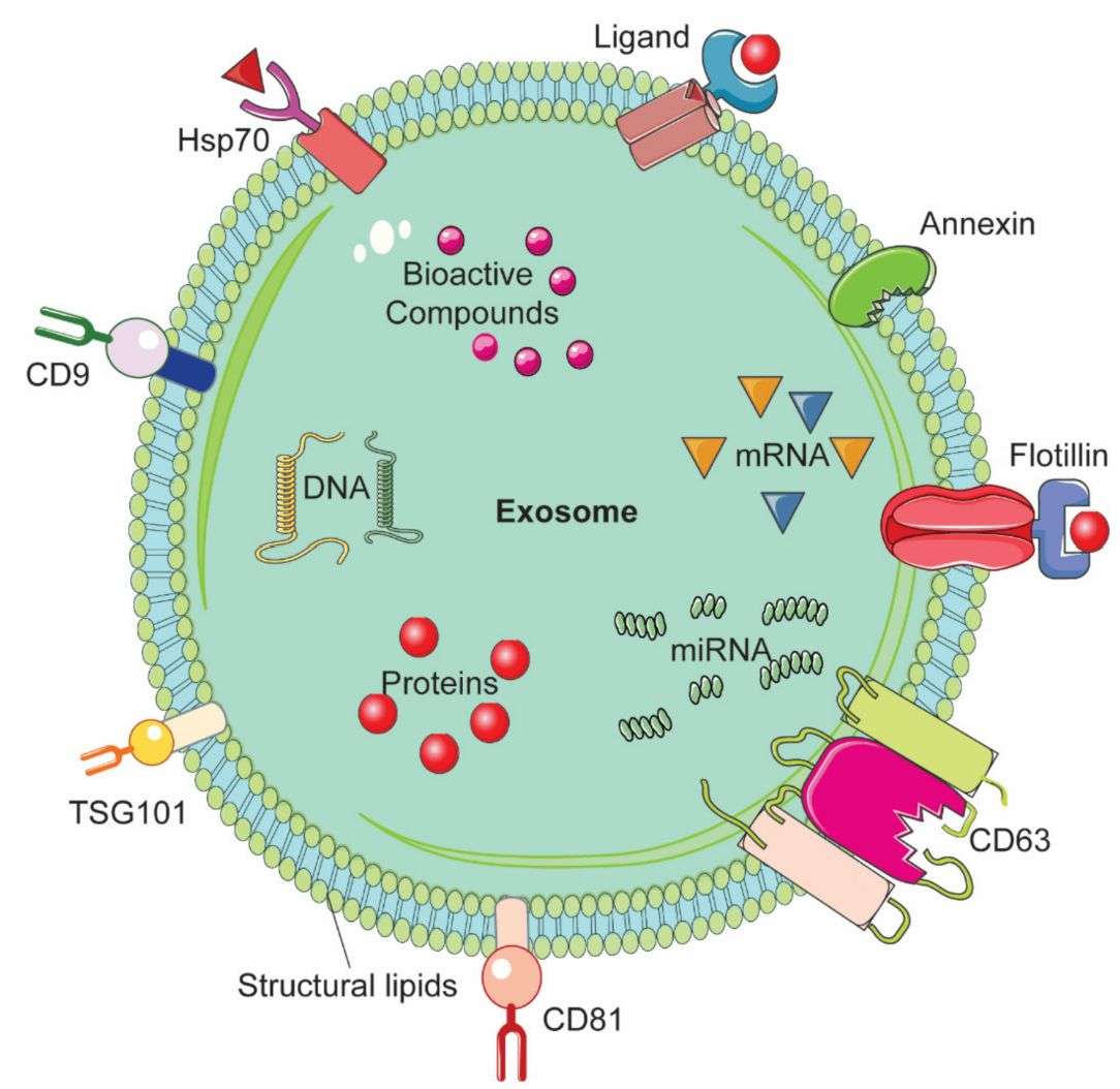
Exosomal RNA fluorescent labeling is the selective staining of RNA cargo enclosed within exosomes using membrane-permeable, RNA-binding fluorophores. These probes typically show low background fluorescence in solution but emit stronger signals after binding to nucleic acids, allowing intraluminal RNA content to be visualized without relying solely on vesicle membrane labeling.
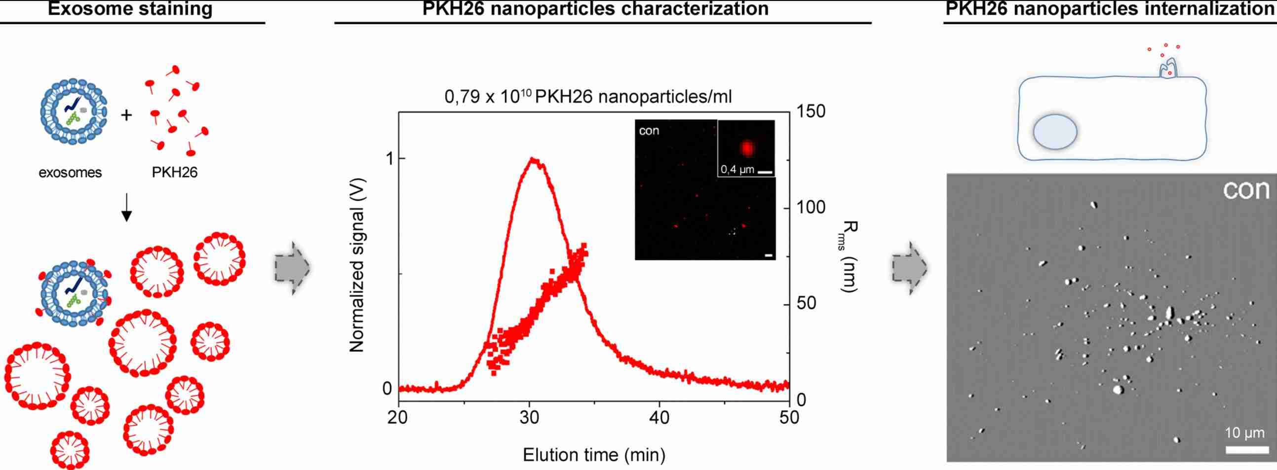
This approach differs fundamentally from lipophilic membrane labeling. While PKH, Di-series, BODIPY-TR ceramide, and related dyes insert into the exosomal lipid bilayer, RNA-selective probes report on internal cargo. This distinction is important for studies that need to differentiate exosome uptake from RNA cargo delivery, particularly because lipophilic dyes may generate non-EV-associated fluorescent particles or transfer between membranes independently of true EV internalization.
RNA-Selective Fluorescent Dyes for Exosome Labeling
| Labeling Option | Principle | Typical Use | Key Notes |
|---|---|---|---|
| RNA-selective green dyes | Penetrate exosomes and fluoresce after binding intraluminal RNA. | General RNA cargo imaging and uptake assays. | Suitable for microscopy, flow cytometry, and dual-labeling workflows. |
| Acridine orange-based dyes | Label nucleic acids inside vesicles with channel-dependent fluorescence. | Multiplex RNA tracking when separated signal channels are needed. | Requires controls to reduce DNA, pH, and free-dye interference. |
| SYTO-family dyes | Provide cell-permeant RNA staining with flexible spectral options. | Multi-color EV labeling and assay compatibility testing. | Dye choice should match filters, autofluorescence, and co-labels. |
| Fluorogenic aptamer systems | Engineered RNA tags activate fluorescence after binding specific dyes. | Sequence-specific tracking of designed RNA cargo. | Best for mechanistic studies requiring RNA engineering. |
| 5-EU metabolic labeling | Labels newly synthesized RNA through metabolic incorporation and click chemistry. | Tracking newly packaged or transferred EV RNA. | Requires donor-cell labeling and EV integrity assessment. |
Our Exosomal RNA Fluorescent Labeling Services
Creative Biostructure offers tiered exosomal RNA fluorescent labeling services to support a wide range of experimental goals, from basic RNA cargo visualization to advanced multiplex tracking. Each project begins with consultation to align the labeling strategy with the exosome source, detection platform, spectral requirements, and biological question.
RNA Dye Portfolio
Our RNA labeling options include:
- RNA-selective green dyes: Suitable for intraluminal RNA staining and compatible with common 488 nm excitation platforms.
- Acridine orange-based dyes: Red-shifted nucleic acid staining options for multiplex experiments requiring separation from green-channel labels.
- SYTO-family dyes: Flexible spectral choices for multi-color EV imaging and assay panel design.
- Fluorogenic RNA aptamer systems: Engineered RNA-labeling approaches for sequence-specific cargo tracking.
- 5-EU metabolic labeling with click chemistry: Bioorthogonal labeling for newly synthesized RNA packaged into EVs.
Labeling Strategies We Offer
- Single-color exosomal RNA staining: Optimization of RNA-selective dye staining, including dye-to-particle ratio, incubation time, and post-labeling purification by size-exclusion chromatography or ultrafiltration to reduce unbound dye.
- Dual-labeling of membrane and RNA cargo: Combination of RNA-selective staining with a spectrally separated membrane dye to compare vesicle uptake with RNA cargo transfer in the same experiment.
- Aptamer-based RNA labeling: Design, expression, and validation of engineered RNA constructs for projects requiring more specific tracking of defined RNA cargo species.
- Metabolic RNA labeling with 5-EU: Donor-cell pulse labeling followed by fluorescent click chemistry after EV isolation, enabling covalent tracking of newly transcribed RNA pools.
- Custom dye validation: Evaluation of new, client-specified, or proprietary RNA-binding fluorophores, including spectral compatibility, dose-response testing, background assessment, and cross-validation with reference staining workflows.
Standard Workflow
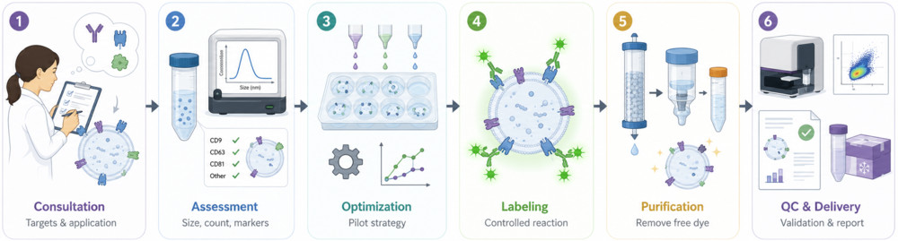
- Project Consultation: Define the exosome source, target RNA cargo, detection platform, spectral channels, and study objectives.
- Baseline Exosome Characterization: Assess particle concentration, size distribution, and key EV markers to confirm sample quality before labeling.
- Dye Selection and Protocol Optimization: Optimize dye type, concentration, incubation conditions, and buffer system to improve RNA labeling efficiency while minimizing background and EV perturbation.
- Controlled RNA Labeling and Purification: Label exosomes under optimized conditions, with optional dual-labeling design, followed by SEC, ultrafiltration, or ultracentrifugation to remove free dye and aggregates.
- Quality Control and Data Delivery: Reassess labeled exosomes, quantify fluorescence signal, perform optional imaging or uptake analysis, and provide a report with methods, QC data, and interpretation.
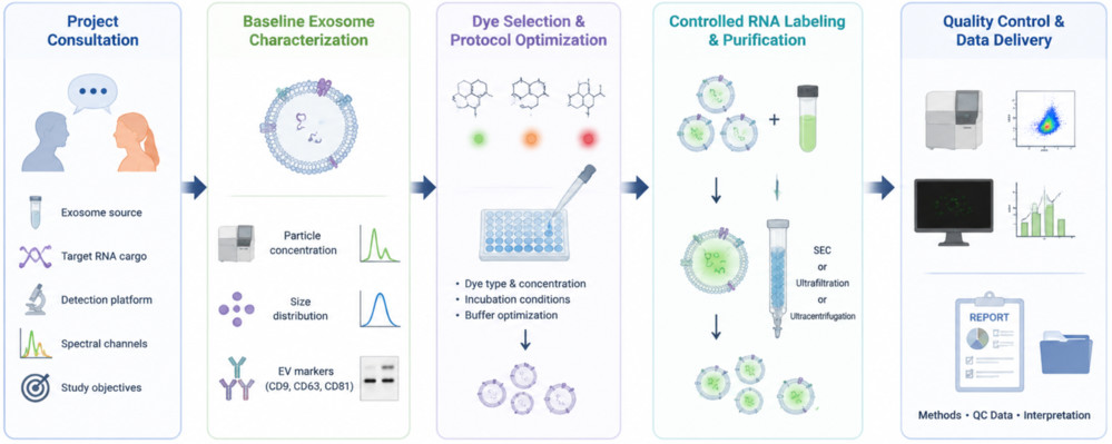 Figure 1. Exosomal RNA Fluorescent Labeling Workflow Overview. (Creative Biostructure)
Comprehensive Quality Control and Characterization
Each labeled exosome batch undergoes a comprehensive quality control workflow to verify labeling performance, EV integrity, and sample purity.
- Particle Characterization: Nanoparticle tracking analysis (NTA) measures particle concentration and size distribution before and after labeling.
- Fluorescence Assessment: Fluorescence intensity and labeling efficiency are quantified using fluorescence spectroscopy, microplate readers, or nano-flow cytometry.
- RNA Integrity: RNA content is evaluated to confirm that the labeling process preserves intraluminal RNA.
- Morphology Analysis: Transmission electron microscopy (TEM) or Cryo-EM verifies exosome morphology and membrane integrity.
- Colloidal Stability: Zeta potential analysis assesses surface charge and suspension stability.
- Purity & Stability (Optional): Free-dye removal is verified using appropriate controls, with optional stability studies to evaluate fluorescence retention and storage performance.
All projects include a comprehensive report summarizing labeling parameters, quality control results, fluorescence data, and supporting documentation. Raw data and imaging files are available upon request.
What Deliverables Will You Receive
| Deliverable | Description |
|---|---|
| Labeled exosome sample | Fluorescently labeled exosomes in optimized buffer, shipped on cold pack or dry ice as appropriate. |
| QC report | Summary of NTA, fluorescence intensity, labeling efficiency, and optional TEM and zeta potential data. |
| Protocol document | Detailed labeling workflow including dye conditions, incubation, purification, and handling guidance. |
| Raw data files | Supporting datasets (e.g., spreadsheets, images, spectra) provided upon request for publication or regulatory use. |
| Technical support | Guidance on data interpretation, imaging setup, and multiplex experiment design. |
Applications
- Exosomal RNA uptake imaging: Visualization of RNA transfer into recipient cells using fluorescence microscopy.
- RNA delivery quantification: Measurement of exosomal RNA delivery efficiency by flow cytometry or nano-flow cytometry, distinguishing true cargo transfer from dye artifacts.
- Intracellular trafficking analysis: Tracking exosome internalization and RNA release dynamics in live cells.
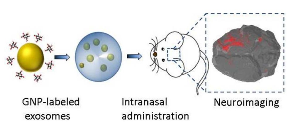
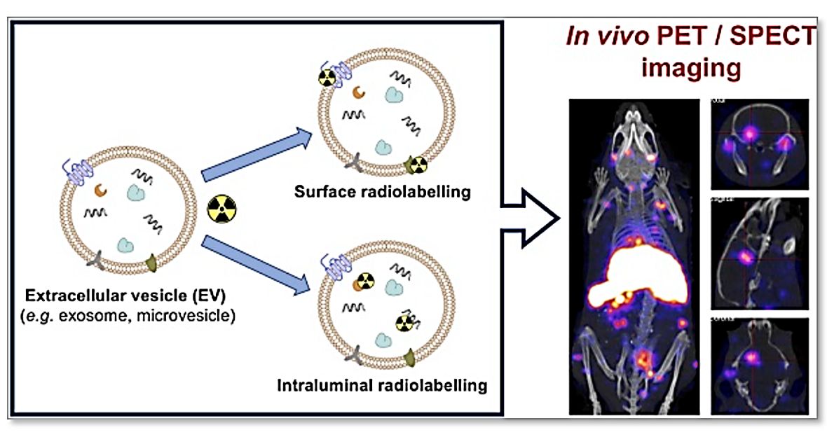
- In vivo biodistribution: Monitoring RNA-loaded exosomes in preclinical models using fluorescence-based or dual-labeling strategies.
- Functional delivery validation: Correlating RNA cargo signal with downstream reporter expression in recipient cells.
- Formulation and QC monitoring: Assessing RNA cargo retention and stability during exosome processing and storage.
How to Get Started
Clients may submit purified exosomes, conditioned media, or biofluid samples. Exosome isolation services are also available as an upstream option for raw biological materials.
Step 1: Provide sample type, target RNA, detection platform, and preferred fluorescence channels.
Step 2: We assess feasibility and recommend an optimized labeling strategy, followed by a tailored technical proposal including workflow, timeline, and deliverables.
Why Choose Creative Biostructure
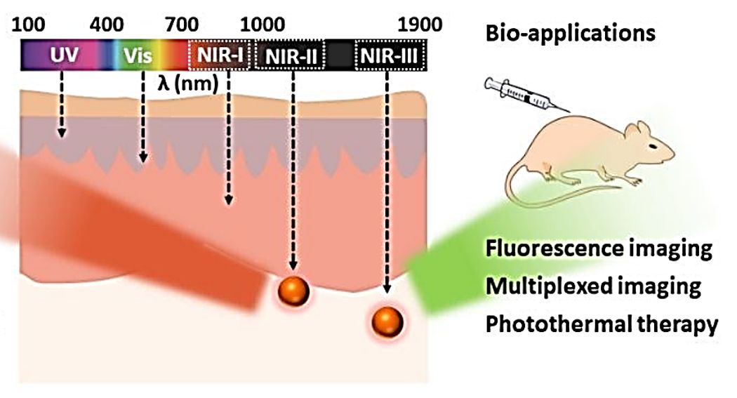
- Exosome labeling expertise: End-to-end experience in exosome engineering and exosome fluorescent labeling across UV to near-infrared spectra.
- High-confidence data quality: Robust controls and nano-flow cytometry workflows reduce dye artifacts and improve EV-specific signal reliability.
- Dual-labeling capability: Simultaneous membrane and RNA cargo tracking to distinguish uptake from true RNA delivery.
- Flexible labeling platforms: RNA-selective dyes, fluorogenic aptamers, and metabolic labeling strategies tailored to project needs.
- Expert scientific support: PhD-level team providing data interpretation and publication-ready documentation.
Case Study
Case: Fluorogenic Aptamer-Based Tracking of Exosomal RNA
Background
Tracking exosomal RNA cargo is difficult because membrane labels mainly show vesicle uptake, not RNA sorting or delivery. This study developed a fluorogenic RNA aptamer system for direct exosomal RNA visualization.
Methods
- Labeling strategy: EXO-Code fused with RNA Mango aptamer
- Fluorogenic dye: Thiazole orange-based signal activation
- Model: Human MSC-derived sEVs
- Analysis: qPCR, NTA, TEM, confocal imaging, live-cell tracking, and single-particle flow cytometry
Results
- EXO-Code sequences showed strong enrichment in sEVs.
- RNA puncta were visualized in cells and vesicles.
- Labeled RNA co-localized with exosome-related markers.
- EXO-Probe-loaded sEVs showed stronger fluorescence than controls.
- Fluorescence signals correlated with NTA-based particle counts.
Conclusion
Fluorogenic aptamer-based labeling enables direct tracking of exosomal RNA sorting, vesicle loading, intracellular movement, and cargo-associated fluorescence, providing a more cargo-focused approach than membrane-only exosome labeling.
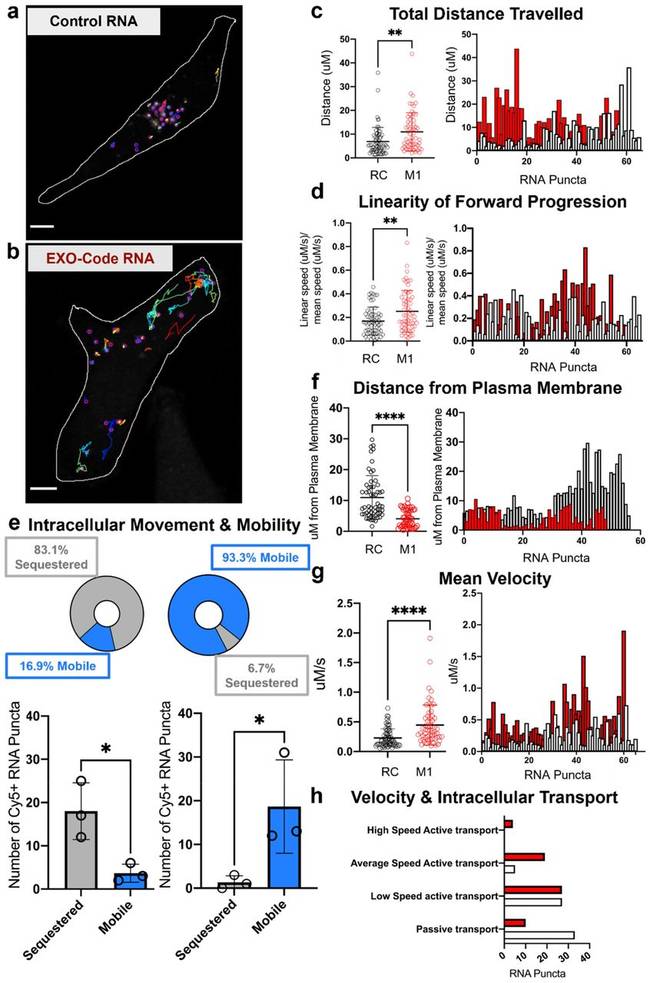 Figure 3. Live-cell imaging of fluorescently labeled EXO-Code RNA in cells, showing intracellular RNA puncta movement, mobility, distance from the plasma membrane, and transport-speed profiles compared with control RNA. (Bonacquisti E E, et al., 2021)
For questions about our Exosomal RNA Fluorescent Labeling Service, to request a quotation, or to discuss a custom labeling project, please submit an inquiry through our online form. Contact us to start your project with a reliable exosomal RNA fluorescent labeling solution.
References
- Manca S, Upadhyaya B, Mutai E, et al. Milk exosomes are bioavailable and distinct microRNA cargos have unique tissue distribution patterns. Scientific Reports. 2018, 8(1): 11321.
- Bonacquisti E E, Ferguson S W, Jasiewicz N E, et al. Fluorogenic EXO-Probe aptamers for imaging and tracking exosomal RNAs. BioRxiv. 2021: 2021.08. 18.456703.
- Bonacquisti E E, Ferguson S W, Wadsworth G M, et al. Fluorogenic RNA-based biomaterials for imaging and tracking the cargo of extracellular vesicles. Journal of Controlled Release. 2024, 374: 349-368.
Frequently Asked Questions
For any inquiries, our support team is ready to help you get technical support for your research and maximize your experience with Creative Biostructure.