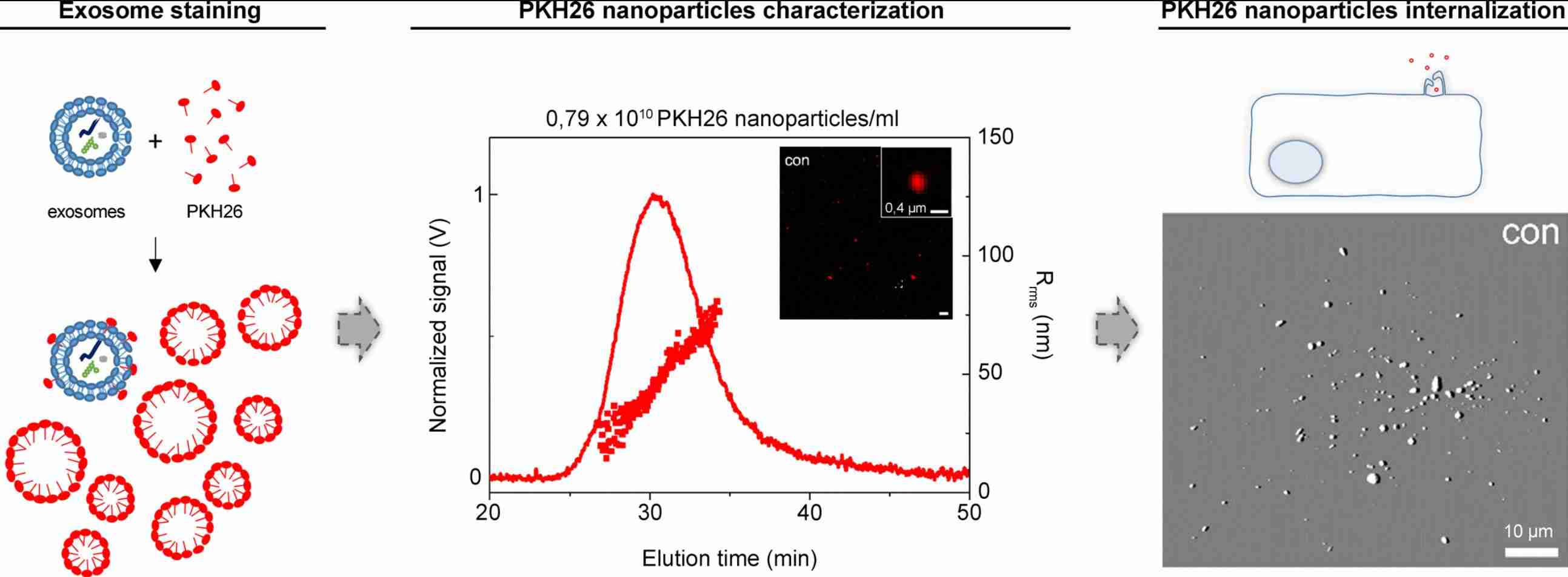
Exosome Labeling with Lipophilic Dyes
Exosome labeling with lipophilic dyes is a widely used, non-genetic approach for tracking extracellular vesicles in uptake, biodistribution, and delivery studies. Creative Biostructure provides optimized lipophilic dye labeling services for exosomes and EVs, using controlled staining conditions, efficient free-dye removal, and rigorous fluorescence and EV characterization to deliver reliable, high signal-to-noise labeled samples for in vitro, in vivo, and preclinical research.
What Is Exosome Labeling with Lipophilic Dyes
Lipophilic dyes are fluorescent small molecules containing long hydrocarbon tails that partition into the phospholipid bilayer of exosomes. Once inserted, the fluorophore headgroup remains exposed to the aqueous environment, enabling detection by fluorescence microscopy, flow cytometry, nano-flow cytometry, and live-animal imaging. This chemical labeling approach does not require transfection or genetic engineering of producer cells, making it compatible with primary cell-derived exosomes, biofluids, and preclinical formulations.
Mechanism of Lipophilic Dye Insertion into the Exosome Membrane
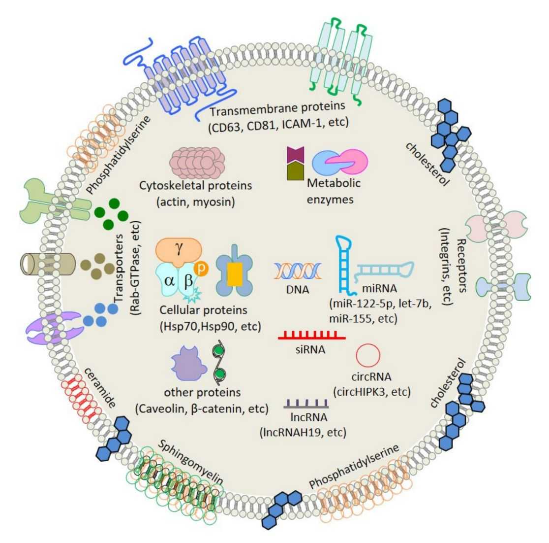
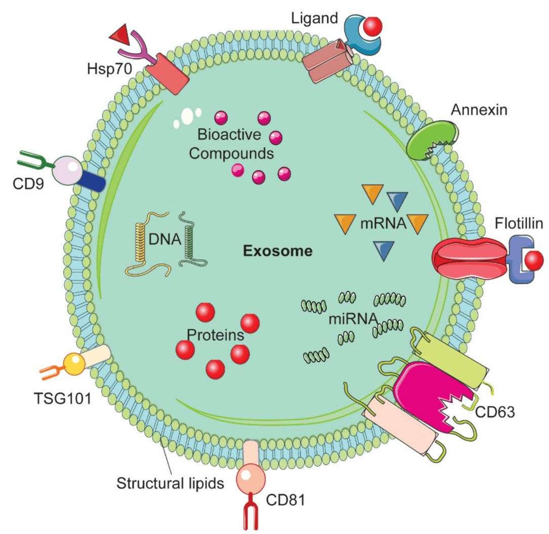
Exosomes possess a cholesterol- and sphingolipid-enriched lipid bilayer with an exposed hydrophobic core. Lipophilic dyes exploit this architecture through a two-step process: (1) the aliphatic tails of the dye intercalate between phospholipid acyl chains, and (2) the fluorophore moiety becomes oriented at the membrane–water interface. In many carbocyanine dyes, fluorescence quantum yield increases markedly in the hydrophobic lipid environment compared with aqueous solution, providing an inherent contrast mechanism. However, because labeling is non-covalent, dyes can also form free-dye aggregates or transfer to lipoproteins, which must be controlled through careful protocol design.
Commonly Used Lipophilic Dye Families
- PKH dyes (PKH26, PKH67): Long-chain aliphatic dyes that insert rapidly into lipid bilayers and are commonly used for cell and exosome tracking. PKH26 emits in the red-orange range (λex/λem ~551/567 nm), while PKH67 emits green (λex/λem ~490/504 nm).
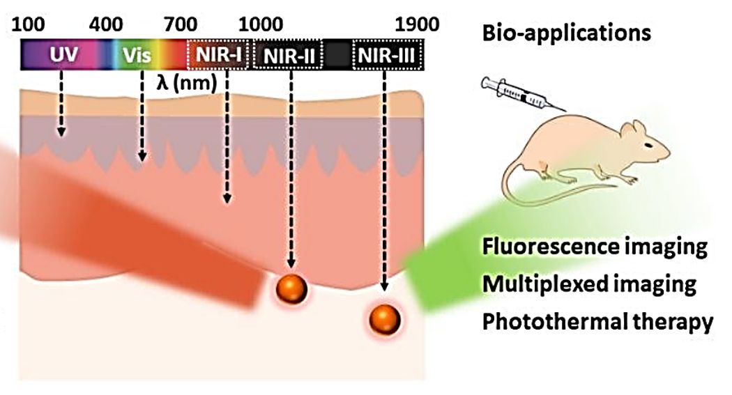
- Carbocyanine dyes (DiO, DiI, DiD, DiR): Long-chain dialkylcarbocyanines that display environment-dependent fluorescence. DiO (green), DiI (orange-red), DiD (red), and DiR (near-infrared, NIR) cover a broad spectral range, with DiR being preferred for in vivo imaging because of reduced tissue autofluorescence.
- BODIPY-TR ceramide: A fluorescent sphingolipid analog used for membrane labeling and dual-labeling experiments with exosomal RNA stains.
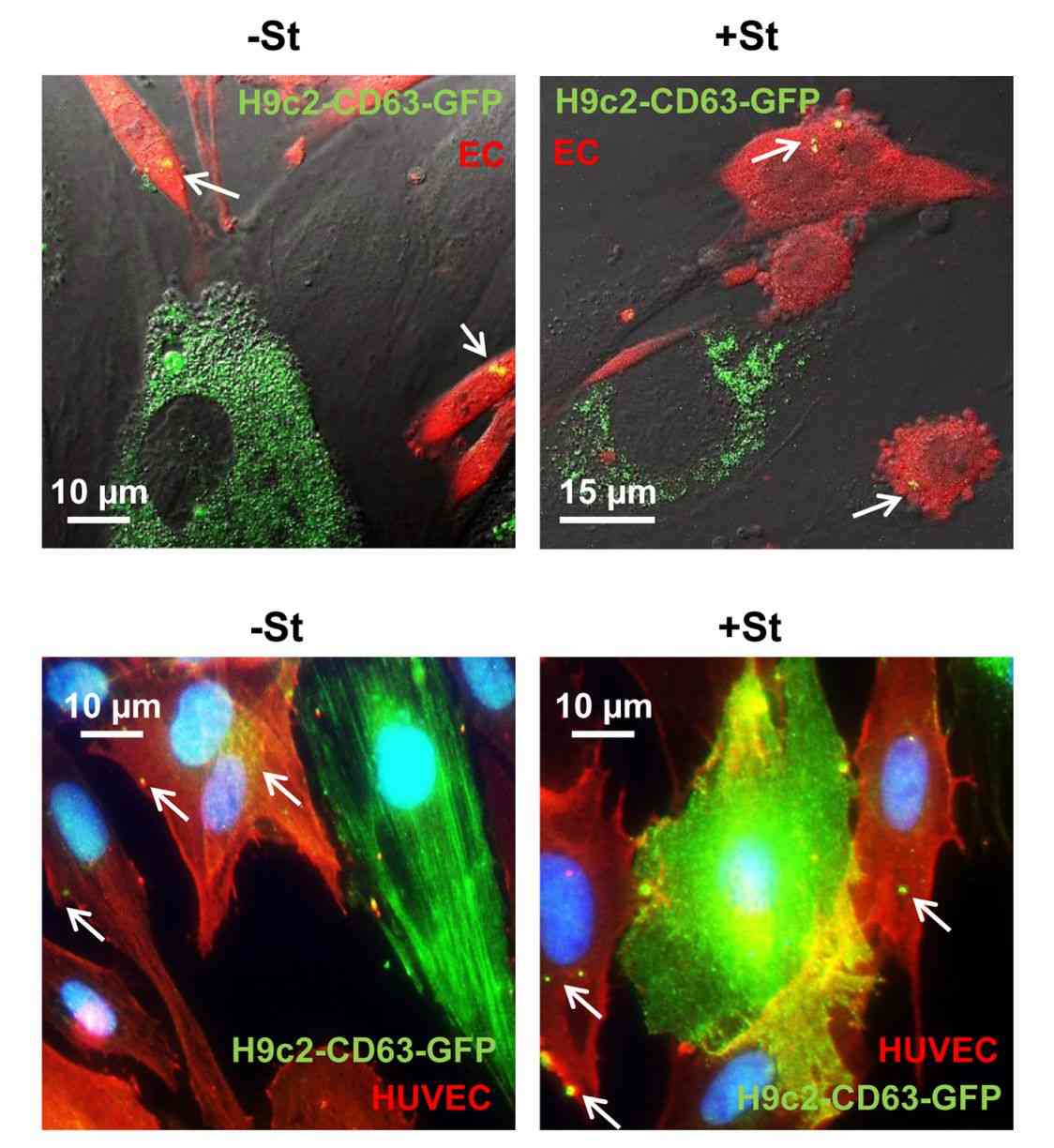
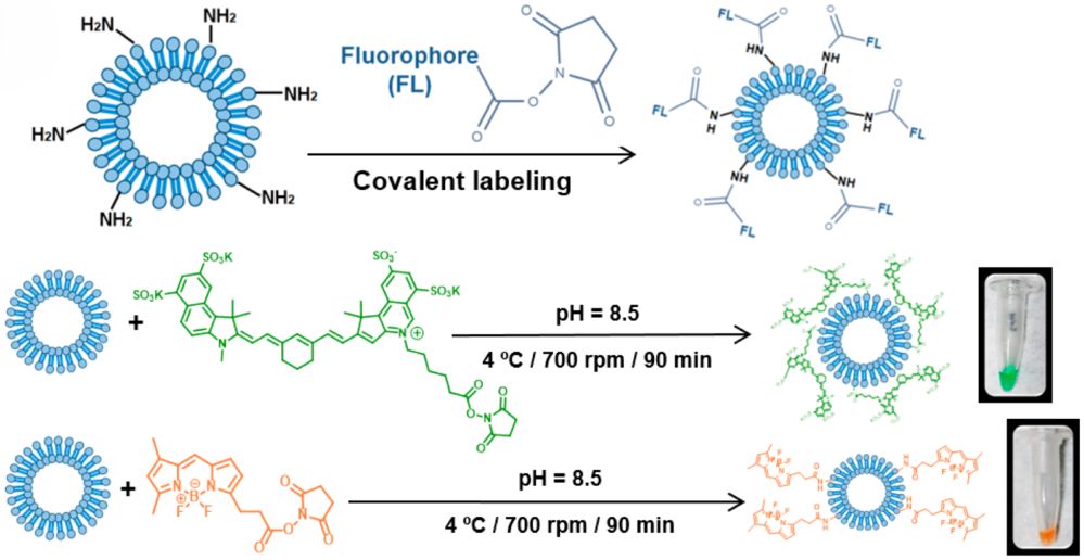 Figure 1. Exosomes labeled with BDP-FL and SCy 7.5 dyes for fluorescent tracking. (González M I, et al., 2021)
Our Advanced Lipophilic Dye Labeling Services
Creative Biostructure provides a comprehensive lipophilic dye labeling service tailored to the research objective—whether the goal is qualitative imaging, quantitative uptake analysis, or preclinical biodistribution tracking. Each project begins with a consultation to match the dye choice, labeling protocol, and purification strategy to the exosome source and downstream readout.
Dye Portfolio and Spectral Options
| Dye | Excitation (nm) | Emission (nm) | Primary Application | Key Considerations |
|---|---|---|---|---|
| PKH67 | 490 | 504 | In vitro uptake and co-localization | Green emission; risk of free-dye aggregates without purification |
| PKH26 | 551 | 567 | In vitro and in vivo tracking | Red-orange emission; extensively validated but requires strict controls |
| DiO | 484 | 501 | Green-channel membrane labeling | Weak fluorescence in water; brightens in lipid environment |
| DiI | 549 | 565 | Red-channel microscopy and flow cytometry | Widely used; aggregation in physiological salt can limit efficiency |
| DiD | 644 | 665 | Far-red multiplexing | Lower autofluorescence background than DiI/PKH26 |
| DiR | 748 | 780 | In vivo NIR imaging | Deep tissue penetration; preferred for whole-animal biodistribution |
| MemGlow-488 | 493 | 515 | Single-EV membrane imaging | Low background; possible passive transfer artifacts |
We select dye pairs for multiplexing experiments based on spectral overlap, brightness, and stability, and we provide single- or dual-color labeling upon request.
Labeling Strategies We Offer
- Conventional direct staining: Optimized PKH and Di-series dye labeling with controlled dye-to-particle ratios and appropriate post-labeling purification, including ultracentrifugation or size-exclusion chromatography (SEC).
- Covalent lipid-based labeling: Amine-reactive or lipid-anchor strategies for applications requiring improved dye retention and reduced signal loss during extended tracking or in vivo circulation.
- Dual-labeling with luminal cargo stains: Combined membrane and intravesicular labeling approaches for parallel tracking of exosome membranes and internal RNA or nucleic acid cargo.
- Custom dye sourcing and validation: Compatibility testing and protocol optimization for proprietary dyes, customized spectra, or project-specific imaging requirements.
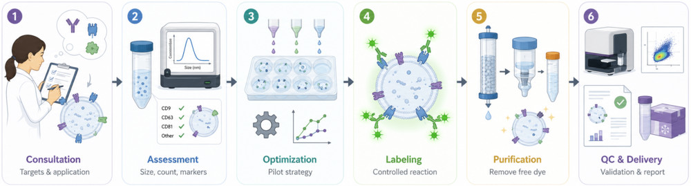
Standard Workflow
- Project Scoping: We define the exosome source, target application, detection method, and preferred dye format.
- Exosome Pre-Assessment: Particle concentration, size distribution, and optional marker profiling are evaluated before labeling.
- Dye & Condition Optimization: Dye type, concentration, incubation time, and buffer conditions are tailored to the sample and downstream assay.
- Controlled Labeling: Exosomes are stained under optimized, low-background conditions to support efficient membrane dye incorporation.
- Purification & Cleanup: Free dye and dye aggregates are removed using validated purification methods such as ultracentrifugation, SEC, or ultrafiltration.
- QC, Reporting & Delivery: Labeled exosomes are assessed by particle and fluorescence analysis, with optional imaging characterization, then delivered with a detailed report and use guidance.
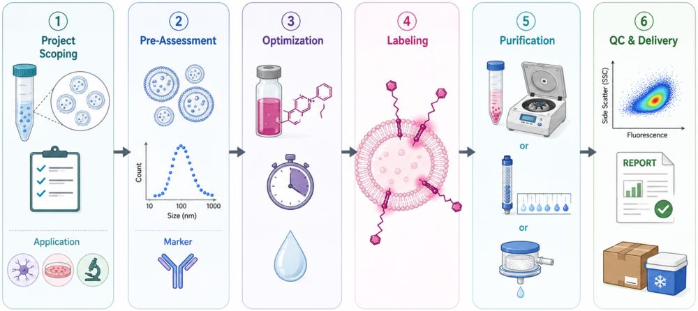 Figure 2. Workflow for Exosome Labeling with Lipophilic Dyes. (Creative Biostructure)
Comprehensive Quality Control and Characterization
Each labeled exosome batch is characterized by a fit-for-purpose analytical package:
- Particle size and concentration (NTA) before and after labeling to detect aggregation or size shifts.
- Fluorescence intensity and labeling efficiency by fluorescence spectrophotometry, plate reader, or nano-flow cytometry.
- Morphology and membrane integrity by Transmission electron microscopy (TEM) or cryo-EM with negative staining.
- Zeta potential to assess surface charge changes induced by dye incorporation.
- Free-dye removal validation using matched dye-only controls and blank purification runs.
- Optional stability testing at 4°C, −20°C, or −80°C to define storage conditions and shelf life.
All results are summarized in a publication-ready report with raw data files available upon request.
Applications of Lipophilic Dye-Labeled Exosomes (Research Use Only)
- In vitro cellular uptake and trafficking studies using confocal microscopy and flow cytometry.
- Single-particle tracking and nano-flow cytometry for high-resolution EV-cell interaction profiling.
- In vivo near-infrared biodistribution and tumor-targeting studies using DiR or NIR covalent labels.
- Co-localization with organelle markers to map exosome intracellular routing after internalization.
- Comparative labeling-method benchmarking to select the optimal tracer for a specific biological question.
- Preclinical screening of engineered exosomes or exosome-mimetic nanovesicles for delivery applications.
How to Get Started
To ensure optimal labeling outcomes, clients may provide isolated exosomes or conditioned medium, or request our exosome isolation service as an upstream add-on. Project initiation follows a simple path:
- Share your exosome source, target dye or spectral channel, and experimental readout.
- Our team evaluates feasibility, recommends dye and protocol options, and provides a customized proposal.
- Upon approval, we execute labeling and characterization under controlled conditions and deliver the final product with full documentation.
What Deliverables Will You Receive
| Deliverable | Description |
|---|---|
| Labeled Exosome Samples | Fluorescently labeled exosomes in your preferred research-grade buffer. |
| Experimental Report | Dye selection, labeling parameters, purification conditions, and recovery data. |
| Characterization Package | NTA size/concentration, fluorescence intensity, TEM or cryo-EM images, and QC summary. |
| Dye-Only Control Data | Control results to support labeling specificity assessment. |
| Optional Add-ons | Stability monitoring, uptake assay, and scale-up recommendations. |
Why Choose Creative Biostructure
- Specialized expertise in exosome engineering, exosome fluorescent labeling, and multi-modal characterization.
- Optimized protocols that address known pitfalls of lipophilic dyes, including free-dye aggregation and buffer-dependent artifacts.
- Flexible dye selection spanning PKH, carbocyanine, NIR, and next-generation membrane probes.
- Scalable workflows from small-scale pilot studies to larger preclinical batches.
- Dedicated scientific support with transparent reporting and publication-ready documentation.
Case Study
Case: Optimized PKH26 Labeling to Reduce Dye Artifacts
Background
Lipophilic dyes such as PKH26 are widely used for EV uptake and biodistribution studies, but non-specific dye particles may cause false-positive signals.
Methods
Small EVs were isolated from osteosarcoma cells and labeled with PKH26 under different quenching conditions, including PBS, 5% dextrose, BSA, and EV-depleted FBS. Labeling outcomes were assessed by NTA, single-EV flow cytometry, TEM, fluorescence imaging, and in vivo lymph node analysis.
Results
Protein-free buffers, especially PBS and 5% dextrose, reduced dye particle formation and improved signal-to-noise ratio. In contrast, protein-containing buffers generated EV-sized dye-protein aggregates that were taken up by cells and detected in vivo, potentially confounding EV tracking results.
Conclusion
Careful buffer selection, dye-only controls, EV purity assessment, and post-labeling QC are essential for reliable lipophilic dye-based exosome tracking.
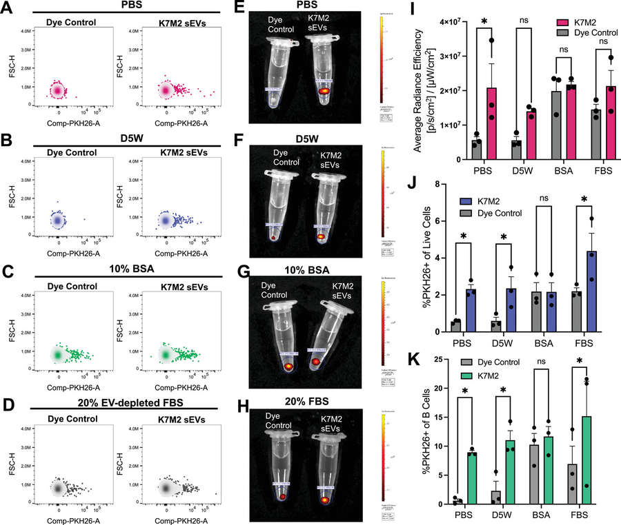 Figure 3. PKH26-labeled sEVs detected in mouse lymph nodes by flow cytometry and IVIS imaging. (Haines L A, et al., 2025)
Ready to generate high-quality, fluorescently labeled exosomes for your imaging, tracking, or delivery project? Contact us to discuss dye selection, labeling strategy, and a customized experimental plan.
References
- González M I, González-Arjona M, Santos-Coquillat A, et al. Covalently labeled fluorescent exosomes for in vitro and in vivo applications. Biomedicines. 2021, 9(1): 81.
- Cha M, Jeong S H, Bae S, et al. Efficient labeling of vesicles with lipophilic fluorescent dyes via the salt-change method. Analytical Chemistry. 2023, 95(14): 5843-5849.
- Chen C, Cai N, Niu Q, et al. Quantitative assessment of lipophilic membrane dye‐based labelling of extracellular vesicles by nano‐flow cytometry. Journal of Extracellular Vesicles. 2023, 12(8): 12351.
- Boudna M, Campos A D, Vychytilova-Faltejskova P, et al. Strategies for labelling of exogenous and endogenous extracellular vesicles and their application for in vitro and in vivo functional studies. Cell Communication and Signaling. 2024, 22(1): 171.
- Haines L A, Baeckler A A, Schofield S J, et al. Non‐Specific Particle Formation During Extracellular Vesicle Labelling With the Lipophilic Membrane Dye PKH26. Journal of Extracellular Vesicles. 2025, 14(5): e70079.
Frequently Asked Questions
For any inquiries, our support team is ready to help you get technical support for your research and maximize your experience with Creative Biostructure.