Exosome Surface Protein Fluorescent Labeling Service
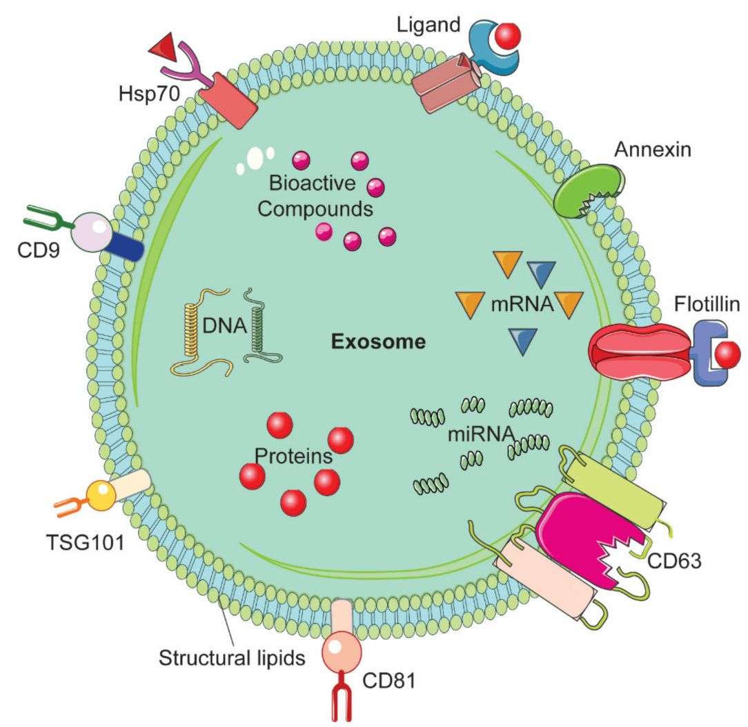
Creative Biostructure provides an Exosome Surface Protein Fluorescent Labeling Service for marker-specific EV detection and characterization. Targeting proteins such as CD9, CD63, CD81, integrins, immunomodulatory ligands, and disease-associated markers, our service enables EV subpopulation analysis and surface proteome profiling with higher molecular specificity than membrane dyes. We combine antibody-based, covalent, bioorthogonal, and aptamer-directed labeling with proper controls and orthogonal validation to deliver reliable, publication-ready data.
What Is Exosome Surface Protein Fluorescent Labeling
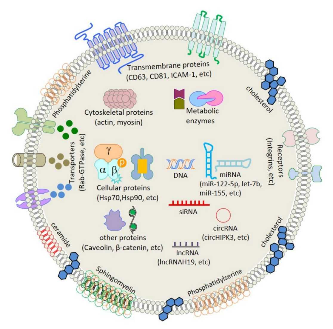
Exosome surface protein fluorescent labeling is a targeted method for attaching fluorophores to proteins exposed on the exosome membrane, enabling marker-specific extracellular vesicle detection and analysis. Unlike lipophilic dyes that stain the lipid bilayer, this approach labels defined surface epitopes, amino acid residues, or glycan structures, producing more specific and stable fluorescence signals. Labeling chemistry directly affects target specificity, signal-to-noise ratio, linkage stability, and compatibility with downstream applications such as flow cytometry, super-resolution microscopy, and single-particle fluorescence imaging.
Why Surface Protein Labeling Over Generic Membrane Staining
Compared with lipophilic dyes such as PKH and Di-series dyes, exosome surface protein fluorescent labeling provides more specific, cleaner, and functionally relevant EV analysis.
- Molecular specificity: Distinguishes EV subpopulations by defined markers such as CD9, CD63, CD81, EpCAM, integrins, or disease-related proteins.
- Reduced artifacts: Lowers non-specific signals, dye-only particles, and dye–protein aggregates commonly seen with membrane dyes.
- Single-EV phenotyping: Supports marker-specific analysis by nano-flow cytometry, super-resolution microscopy, and single-particle imaging.
- Functional preservation: Uses mild aqueous conditions to better retain uptake, immune interaction, and targeting functions.
- Versatile detection: Compatible with microscopy, flow/nano-flow cytometry, plate-reader assays, and dual-compartment tracking.
Labeling Modality Comparison
| Modality | Target Specificity | Label Stability | Throughput | Best For | Limitations |
|---|---|---|---|---|---|
| Antibody (direct) | High (epitope-specific) | Moderate | High (bulk + single-EV) | Routine phenotyping, flow cytometry | Antibody cost; steric hindrance on small EVs |
| Covalent (NHS ester) | Moderate (amine-reactive) | Very high (covalent) | High | In vivo tracking, NIR imaging | Non-specific labeling of non-EV proteins |
| Bioorthogonal click | Selective (nascent EVs) | Very high (covalent) | Moderate | Pulse-chase, in vivo tracking | Requires producer-cell pre-treatment |
| Aptamer | High (target-specific) | Moderate–high | High | Biosensor integration, multiplex panels | Nuclease susceptibility; fewer validated targets |
| Enzymatic/Proximity | Very high (site-specific) | High (covalent) | Low-moderate | Single-EV super-resolution imaging | Requires genetic modification or specialized reagents |
Our Surface Protein Fluorescent Labeling Services
Creative Biostructure delivers custom surface protein labeling solutions across all five modalities described above. Our service is modular: clients may select a single labeling approach or combine complementary methods within a single project.
Fluorescent Probe Portfolio
We stock validated fluorophores across the full visible-to-NIR spectrum for surface protein conjugation:
| Fluorophore Class | Examples | Ex/Em Range (nm) | Conjugation Chemistry | Typical Application |
|---|---|---|---|---|
| CF Dyes | CF488A, CF568, CF660R | 490–660 / 515–680 | NHS ester, click chemistry | Super-resolution (STORM, PALM) |
| ATTO Dyes | ATTO 488, ATTO 647N | 501–646 / 523–664 | NHS ester, maleimide | Single-molecule imaging |
| Cyanine | Cy3, Cy5, Cy7 | 550–750 / 570–773 | NHS ester, DBCO-click | NIR in vivo imaging |
| Tandem Dyes | PE-Cy5, APC-Cy7 | 488/633 ex; 670/785 em | Antibody pre-conjugated | Multicolor flow cytometry panels |
| Qdot | QD525, QD655, QD800 | UV/405 ex; 525–800 em | Streptavidin–biotin | Multiplex single-EV phenotyping |
Service Packages
Package 1 — Targeted Immunophenotyping
- Fluorescent antibody labeling of up to three exosome surface markers.
- Includes antibody titration, isotype controls, and compensation controls.
- Nano-flow cytometry reports marker-positive EVs, MFI, and particle concentration.
- Recommended for: EV subpopulation profiling, biomarker validation, and disease-EV immunophenotyping.
Package 2 — Covalent NHS Ester Protein Labeling
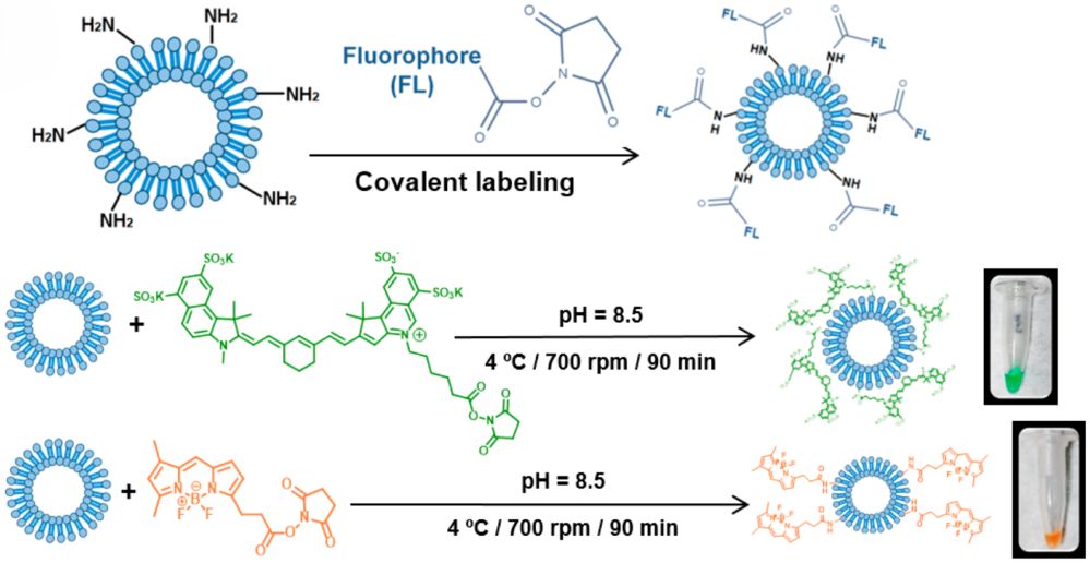
- Covalent labeling of exosome surface proteins with AF647, Cy5, Cy7, BDP-FL, or client-supplied dyes.
- Optimized dye-to-protein ratio with NTA, zeta potential, and nano-flow cytometry QC.
- SEC or ultracentrifugation removes free dye; dye-only controls verify background.
- Recommended for: In vivo biodistribution, long-term EV tracking, and NIR imaging.
Package 3 — Bioorthogonal Glycan/Azide-Click Labeling
- Metabolic Ac4ManNAz labeling followed by EV isolation and DBCO-fluorophore click conjugation.
- Includes azide verification and labeling-efficiency assessment.
- Optional GPI-anchor azide labeling supports alternative EV tracking.
- Recommended for: Pulse-chase studies, in vivo fate mapping, and EV dynamics analysis.
Package 4 — Aptamer Synthesis & Labeling
- Custom DNA aptamer design against client-specified exosome surface targets.
- 5' or 3' fluorophore/biotin conjugation with optional nuclease-resistant modifications.
- Binding and labeling performance validated by SPR, MST, or EV-specific optimization.
- Recommended for: Biosensors, aptamer-based EV capture, and antibody-free detection.
Package 5 — Site-Specific Enzymatic Labeling
- Optional proximity biotinylation enables native exosome surface labeling.
- Single-EV fluorescence validated by super-resolution or interferometric imaging.
- Recommended for: Single-EV protein mapping, stoichiometric labeling, and native EV functionalization.
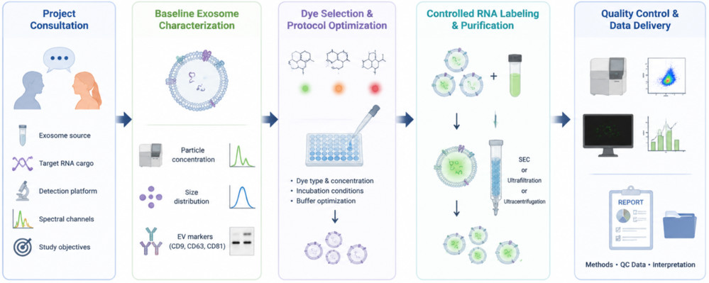
Standard Workflow
- Consultation & Target Definition: Define target proteins, labeling method, fluorophore, sample type, and application.
- Sample Assessment: Evaluate EV size, concentration, and marker expression by NTA and optional protein analysis.
- Strategy Optimization: Select and optimize the most suitable labeling approach at pilot scale.
- Controlled Labeling: Perform full-scale fluorescent labeling with matched experimental controls.
- Purification: Remove free dye, antibodies, or residual reagents by SEC, ultrafiltration, or ultracentrifugation.
- QC & Delivery: Verify EV integrity, labeling efficiency, and fluorescence performance, then deliver labeled EVs with a QC report.
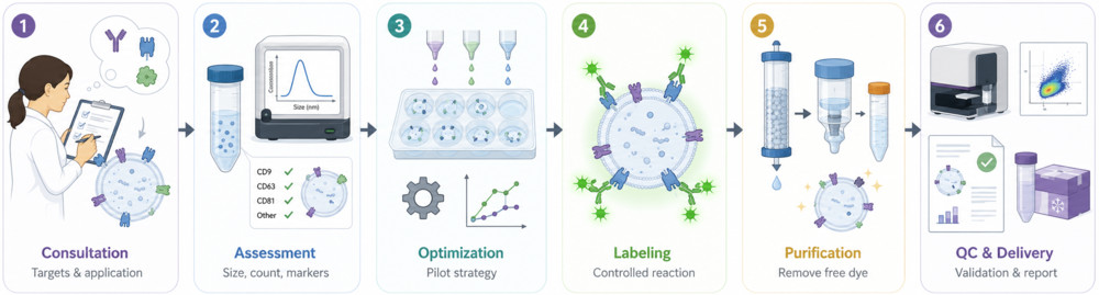 Figure 1. Exosome Surface Protein Fluorescent Labeling Workflow. (Creative Biostructure)
Quality Control and Characterization Standards
Each labeled exosome batch must pass defined QC criteria before release:
- Size and concentration: NTA compares pre- and post-labeling profiles to detect aggregation or vesicle disruption.
- Labeling efficiency: Nano-flow cytometry or single-particle fluorescence imaging measures fluorescent-positive EVs and MFI.
- Specificity: Isotype, scrambled aptamer, or dye-only controls confirm low background signal.
- Morphology: Transmission electron microscopy (TEM) or Cryo-EM verifies intact vesicle structure after labeling.
- Surface integrity: Zeta potential changes are monitored to assess surface charge stability.
- Free-probe removal: Blank purification runs and fluorescence quantification confirm removal of residual dye or probe.
- Stability testing: Optional storage studies assess fluorescence retention at 4°C, −20°C, or −80°C.
All results are summarized in a publication-ready QC report, with raw data available upon request.
Applications (Research Use Only)
- Single-EV immunophenotyping: Identify CD9+, CD63+, and CD81+ EV subpopulations and link marker profiles to cell origin, disease status, or treatment response.
- Multiplex EV profiling: Analyze multiple EV surface epitopes using bead-based fluorescent antibody panels.
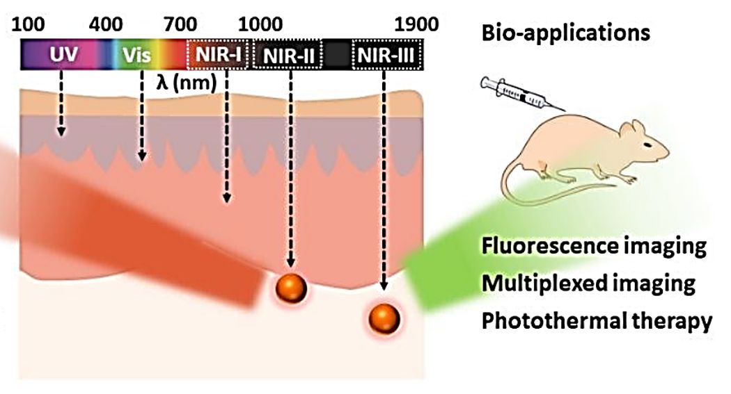
- In vivo NIR biodistribution: Track labeled exosomes by whole-animal fluorescence imaging and organ-level quantification.
- Super-resolution protein mapping: Visualize nanoscale protein distribution on individual exosomes by STORM, PALM, or related imaging methods.
- Aptamer-based EV biosensors: Support fluorescent aptamer probes for microfluidic, SPR, or electrochemical EV detection.
- Pulse-chase EV dynamics: Track nascent EVs and monitor secretion, surface proteome changes, and tissue distribution.
- Engineered EV QC: Verify targeting ligand retention and orientation on drug-delivery exosomes.
How to Get Started
We accept isolated exosomes, conditioned medium, or biofluids, with optional upstream support.
- Submit EV source, target surface protein(s), labeling modality, fluorophore(s), sample amount, and downstream readout through our inquiry form.
- Our scientific team reviews feasibility and provides a tailored strategy, timeline, and quotation.
- Upon approval, we perform labeling, provide progress updates, and deliver labeled EVs with a QC report and raw data files.
What Deliverables Will You Receive
| Deliverable | Description |
|---|---|
| Labeled Exosome Sample | Fluorescently labeled exosomes in an application-compatible research buffer. |
| QC Report | NTA, labeling efficiency, MFI, TEM/cryo-EM, zeta potential, and specificity control data. |
| Method Details | Labeling chemistry, reagent lots, reaction conditions, and purification parameters. |
| Control Data | Isotype, scrambled, dye-only, or azide-free controls based on the workflow. |
| Optional Data | Stability results, scale-up advice, and downstream assay guidance. |
Why Choose Creative Biostructure
- Multi-modality expertise: We match each project with the most suitable labeling strategy, including antibody labeling, NHS ester conjugation, bioorthogonal click chemistry, aptamer labeling, and enzymatic modification.
- MISEV-aligned rigor: Each workflow includes proper specificity controls, orthogonal EV characterization, and transparent reporting for reliable surface protein analysis.
- Single-particle resolution: Nano-flow cytometry, single-particle imaging, and super-resolution microscopy support quantitative, vesicle-level fluorescence assessment.
- Literature-informed protocols: Our methods are benchmarked against peer-reviewed EV labeling studies to reduce technical risk.
Case Study
Case: Covalent Fluorescent Exosome Labeling for In Vitro and In Vivo Tracking
Background
Stable fluorescent labeling is critical for studying exosome uptake, biodistribution, and drug delivery behavior. This study used covalent dye attachment to reduce leakage and false signals associated with lipophilic membrane dyes.
Methods
- Labeling strategy: Succinimidyl ester fluorophores conjugated to exosome surface amine groups
- Fluorophores: BDP-FL and sulfo-cyanine 7.5
- Exosome sources: Goat milk, U87, and B16F10 cells
- Analysis: Flow cytometry, TEM, DLS/NTA, HPLC, confocal microscopy, and in vivo optical imaging
Results
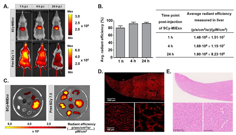 Figure 2. Near-infrared imaging shows the biodistribution of SCy 7.5-labeled milk exosomes in mice, with liver-dominant accumulation confirmed by ex vivo organ imaging and liver-section fluorescence analysis. Free dye displayed a distinct distribution pattern, supporting the stability of covalent exosome labeling. (González M I, et al., 2021)
- Efficient labeling: Over 99% fluorescent-positive exosome events were detected.
- Preserved properties: Labeled exosomes retained morphology and size profiles.
- Improved stability: HPLC showed minimal free-dye release for BDP-FL-labeled exosomes.
- Cellular uptake: SCy 7.5-labeled exosomes entered hepatocytes in a dose- and time-dependent manner.
- In vivo tracking: NIR-labeled exosomes showed biodistribution distinct from free dye.
Conclusion
Covalent exosome surface fluorescent labeling generates stable EV probes for cellular uptake, biodistribution, and optical imaging studies.
Ready to advance your exosome research with molecularly specific surface protein fluorescent labeling? Contact us to discuss your target proteins, labeling modality, and experimental design. We deliver publication-grade labeled EVs with full characterization and transparent reporting.
References
- González M I, González-Arjona M, Santos-Coquillat A, et al. Covalently labeled fluorescent exosomes for in vitro and in vivo applications. Biomedicines. 2021, 9(1): 81.
- Wu Q, Wang W, Zhang C, et al. Capturing nascent extracellular vesicles by metabolic glycan labeling-assisted microfluidics. Nature Communications. 2023, 14(1): 6541.
- Ueda Y, Manabe Y, Saito S, et al. Antibody-Guided Proximity Labeling Enables Selective Chemical Modification of Exosome Surface Proteins. 2026.
Frequently Asked Questions
For any inquiries, our support team is ready to help you get technical support for your research and maximize your experience with Creative Biostructure.