Exosome Labeling with Gold Nanoparticles
Gold nanoparticles (GNPs) are widely used for exosome labeling due to their strong electron density for X-ray and electron microscopy contrast, tunable localized surface plasmon resonance (LSPR) enabling optical, photoacoustic, and photothermal applications, and highly modifiable surface chemistry for targeted functionalization and cargo loading. At Creative Biostructure, our service provides optimized GNP-EV conjugation, rigorous purification, and multi-modal characterization to generate high-quality, publication-ready labeled exosomes for imaging, biodistribution, and therapeutic studies.
What Is Exosome Labeling with Gold Nanoparticles
Exosome labeling with gold nanoparticles is the conjugation of nanoscale gold particles (spherical, rod, or star-shaped) with extracellular vesicles to enable high-contrast tracking based on their strong plasmonic, electronic, and optical properties. GNP-labeled exosomes provide superior stability compared to fluorescent dyes and generate strong signals for TEM, CT, dark-field microscopy, SERS, and photothermal imaging, making them widely used for biodistribution analysis, targeting validation, and theranostic applications.
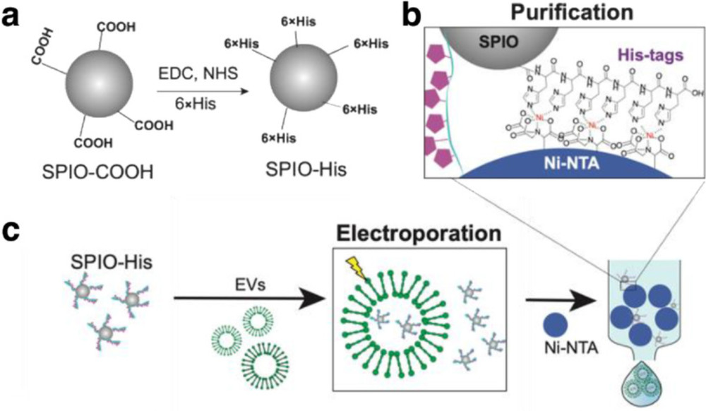
GNP loading is mainly achieved through three strategies: membrane adsorption/insertion using surface-modified GNPs for lipid bilayer association and in vivo tracking; active loading via electroporation or membrane permeabilization for intraluminal encapsulation in SERS or photothermal studies while preserving EV integrity; and producer-cell loading, where GNPs are endocytosed and packaged into secreted exosomes, enabling stable in vivo imaging with reduced background signals.
Our Advanced Gold Nanoparticle Exosome Labeling Services
We offer comprehensive gold nanoparticle exosome labeling for biodistribution studies and preclinical theranostic development. Each project begins with a technical consultation to define the optimal GNP design, surface chemistry, and loading strategy based on the exosome source and intended imaging or therapeutic application.
Gold Nanoparticle Portfolio and Application Matrix
We stock validated fluorophores across the full visible-to-NIR spectrum for surface protein conjugation:
| GNP Type | Typical Size | LSPR Peak | Primary Imaging Modality | Key Considerations |
|---|---|---|---|---|
| Spherical (AuNS) | 5-60 nm | 510-530 nm | CT, TEM, dark-field | High X-ray attenuation; size overlaps with sEV range |
| Gold nanorods (AuNR) | 10×40 nm, aspect 3-4 | 700-850 nm (NIR-I) | Dark-field, photothermal, PA imaging | Strong longitudinal LSPR; sensitive to aspect ratio |
| Gold nanostars (AuNSt) | 30-80 nm | 700-900 nm | SERS, photothermal | High tip field enhancement; excellent SERS substrates |
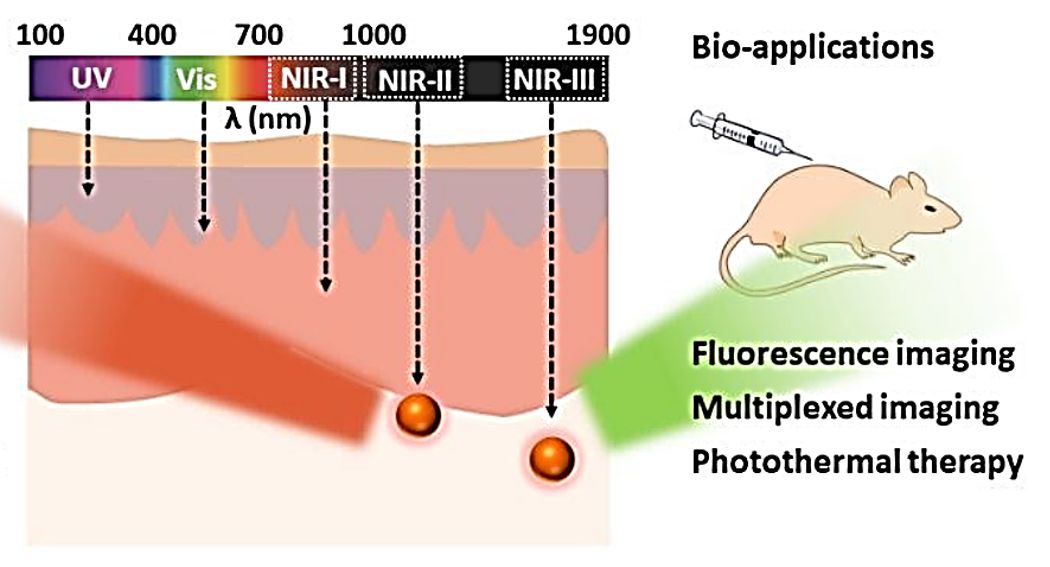
| Gold nanocages / hollow shells | 40-80 nm | 700-1100 nm | NIR-II photothermal, photoacoustic | Tunable LSPR across NIR window |
| Glucose-coated GNP | 10-50 nm | 510-530 nm | CT, biosynthetic EV loading | GLUT-1 mediated uptake; biosynthetic packaging |
| PEG/peptide-functionalized GNP | 20-80 nm | Tunable | Targeted delivery, theranostics | Custom ligand density; reduced RES clearance |
Geometry, LSPR peak, and surface functionalization are matched to the intended imaging modality, and we provide custom architectures on request.
Labeling Strategies We Offer
We provide multiple gold nanoparticle (GNP)-exosome labeling strategies selected based on imaging modality, loading location, and study purpose:
- Surface adsorption/insertion: PEGylated or citrate-stabilized GNPs associate with the exosome membrane under controlled conditions, enabling rapid labeling for in vitro studies and short-term in vivo tracking.
- Active intraluminal loading: Electroporation or mild membrane permeabilization allows GNP entry into exosomes while preserving vesicle integrity, suitable for high-sensitivity imaging applications (e.g., SERS, photothermal readouts).
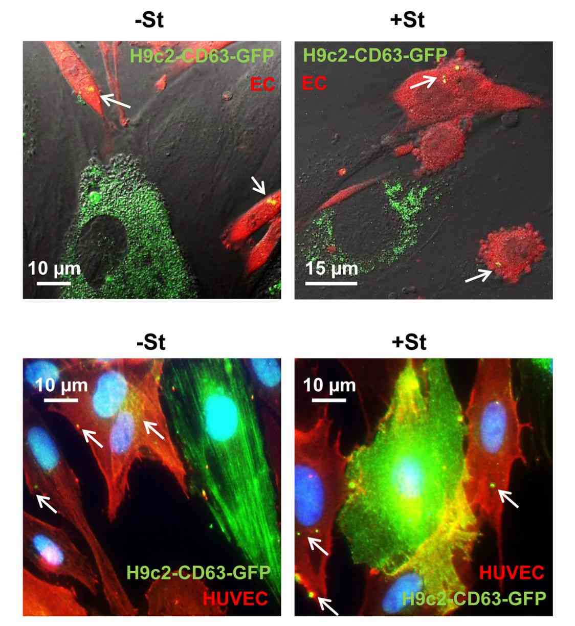
- Producer-cell loading: Functionalized GNPs are internalized by donor cells and incorporated into secreted exosomes, providing stable, biologically derived labeling with reduced background for in vivo imaging.
- Surface covalent conjugation: Chemistries such as thiol-based coupling or click reactions anchor GNPs to exosomal surface proteins for stable, targetable constructs in biodistribution and theranostic studies.
- Custom GNP-EV design: Tailored nanoparticle size, shape, and surface ligands are engineered to meet specific imaging, targeting, or translational requirements, with full characterization and stability validation.
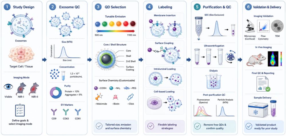
Standard Workflow
- Project Design: Define exosome source, target, and imaging/therapeutic goal to select the optimal GNP strategy.
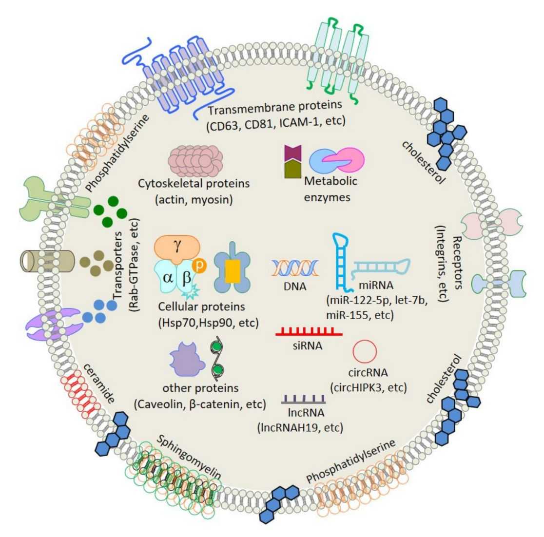
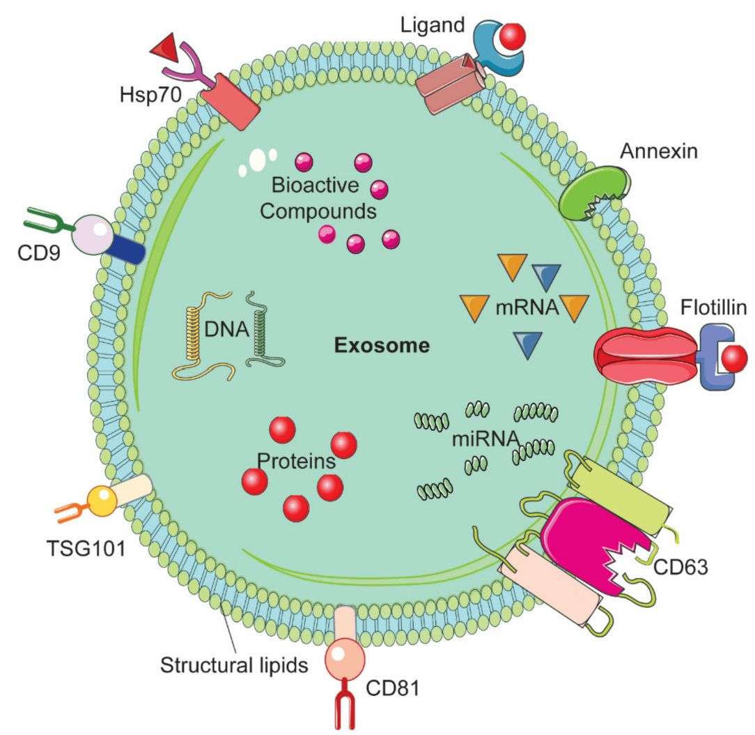
- Exosome QC: Verify size, concentration, and key markers (CD9/CD63/CD81).
- GNP Preparation: Select or customize nanoparticle size, shape, and surface chemistry.
- Labeling: Perform optimized GNP-exosome conjugation (surface, intraluminal, or biosynthetic).
- Purification & QC: Remove free GNPs and confirm purity via physicochemical characterization.
- Validation & Delivery: Validate labeling by TEM/spectroscopy/imaging and deliver with full data package.
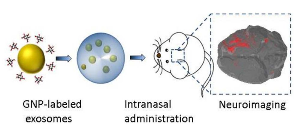
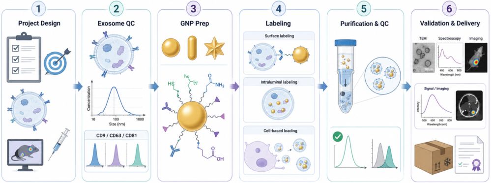 Figure 1. Gold Nanoparticle Exosome Labeling Workflow for Imaging. (Creative Biostructure)
Comprehensive Quality Control and Characterization
Each GNP-labeled exosome batch includes a streamlined QC package focused on key performance and identity parameters:
- Size & stability: NTA/DLS before and after labeling to confirm integrity
- GNP loading: ICP-MS (gold per EV) with UV-Vis (LSPR) confirmation
- Surface charge: Zeta potential in working buffer
- Structure: Transmission electron microscopy (TEM) or Cryo-EM for morphology and GNP association
- EV identity: CD9/CD63/CD81 retention by nano-flow cytometry or WB
- Purity: Free-GNP removal verified via matched controls
- Optional validation: In vitro uptake and in vivo imaging (CT, SERS, photothermal)
All datasets are delivered in a publication-ready report with raw data for downstream use.
What Deliverables Will You Receive
| Category | Deliverable | Description |
|---|---|---|
| Final Product | GNP-labeled exosomes | Research-grade exosomes in optimized buffer with defined storage conditions |
| Core Documentation | Technical project report | GNP design rationale, labeling strategy, purification workflow, and yield summary |
| Characterization Data | Multi-modal QC dataset | NTA, DLS, zeta potential, UV-Vis (LSPR), ICP-MS (gold quantification), TEM/cryo-EM |
| Specificity Controls | Free-GNP & matched controls | Background and specificity validation for imaging and biodistribution studies |
| Optional Add-ons | Functional validation & scale guidance | CT/dark-field/SERS/photothermal testing and scale-up recommendations if required |
Applications of Gold Nanoparticle-Labeled Exosomes (Research Use Only)
GNP-labeled exosomes enable high-contrast, multi-modal tracking and functional studies across preclinical research:
- In vivo biodistribution imaging: CT-based tracking of exosome distribution in tumor, cardiac injury, and neurodegenerative models.
- Deep-tissue imaging: Dark-field and photoacoustic imaging leveraging tunable LSPR for enhanced tissue penetration and contrast.
- Molecular profiling: SERS-based multiplex detection of exosome surface markers and cargo.
- Ultrastructural localization: Cryo-EM and CLEM for high-resolution tracking of exosomes in cells and tissues.
How to Get Started
Project initiation is designed to be fast and streamlined. Clients may provide isolated exosomes, conditioned medium, or producer cells depending on the selected labeling strategy.
- Share requirements: exosome source, target tissue/cell, and intended imaging.
- Feasibility review: we assess compatibility and recommend the optimal GNP design and labeling strategy.
Why Choose Creative Biostructure
- Multi-architecture capability: spherical, rod, star, cage, and glucose-functionalized gold nanoparticles integrated into a single standardized workflow.
- Application-aligned loading strategies: from passive adsorption to biosynthetic GLUT-1–mediated packaging, optimized for specific imaging.
- Comprehensive QC package: including ICP-MS gold quantification, exosome surface-marker profiling, and matched free-GNP controls for reliability and specificity.
- Scalable & publication-ready workflows: from pilot studies to preclinical batches with consistent reproducibility, transparent reporting, and full documentation.
Case Study
Case: CT Tracking of GNP-Labeled Exosomes in Myocardial Infarction
Background
Native exosomes are difficult to monitor by CT. This study used gold nanoparticles (GNPs) to label MSC-derived exosomes for non-invasive tracking in a myocardial infarction mouse model.
Methods
- Labeling strategy: Glucose-modified GNPs
- Exosome source: MSC-derived exosomes
- Model: Myocardial infarction mice
- Tracking focus: Retention and biodistribution after intramyocardial injection
- Analysis: NTA, TEM, DLS/zeta potential, density-gradient purification, and CT imaging
Results
- Efficient labeling: GNPs were successfully loaded into exosomes.
- Maintained integrity: NTA showed no obvious aggregation after labeling.
- Confirmed localization: TEM verified GNP association with exosomes.
- Reduced background: Density-gradient purification removed excess free GNPs.
- CT-trackable signal: Labeled exosomes were visible in the infarcted myocardium at 4 h and 24 h.
- Localized retention: Most signals remained near the injury site, with limited distribution to other organs.
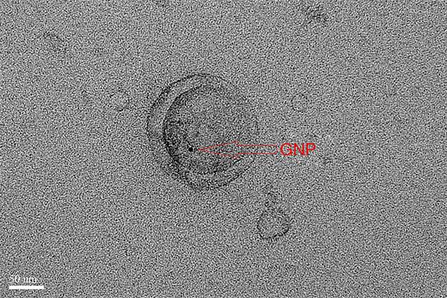 Figure 2. Transmission electron microscopy (TEM) showing exosomes incubated with gold nanoparticles (GNPs) at 37 °C for 10 h. (Gong L, et al., 2021)
Conclusion
GNP labeling enables CT-compatible exosome tracking in deep tissues, supporting its use in biodistribution, pharmacokinetic, and preclinical exosome delivery studies.
Ready to generate high-quality gold nanoparticle-labeled exosomes for your imaging, biodistribution, or theranostic project? Contact us to discuss GNP architecture, loading strategy, and a customized experimental plan.
References
- Betzer O, Perets N, Angel A, et al. In vivo neuroimaging of exosomes using gold nanoparticles. ACS nano. 2017, 11(11): 10883-10893.
- Gong L, Weng Y, Zhou W, et al. In vivo CT imaging of gold nanoparticle-labeled exosomes in a myocardial infarction mouse model. Annals of Translational Medicine. 2021, 9(6): 504.
Frequently Asked Questions
For any inquiries, our support team is ready to help you get technical support for your research and maximize your experience with Creative Biostructure.