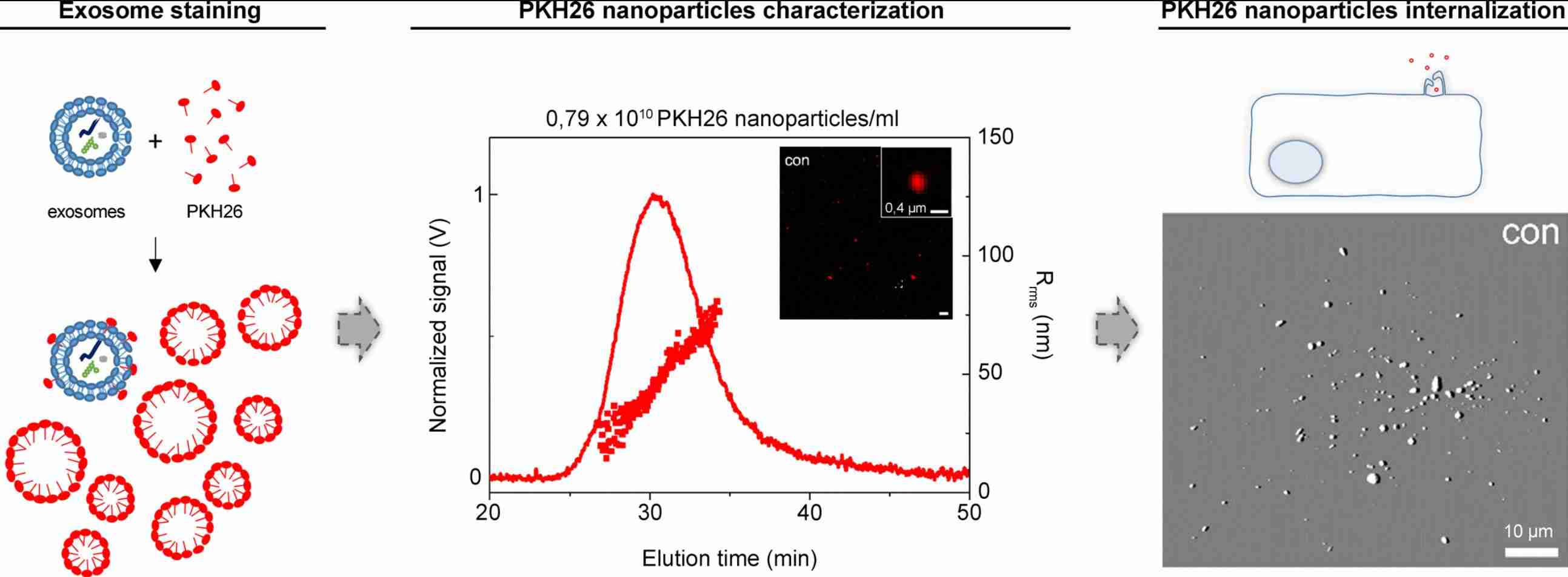
Exosome Labeling with SPION for MRI Tracking
Superparamagnetic iron oxide nanoparticles (SPIONs) enable high-resolution MRI-based exosome imaging with deep tissue penetration and no ionizing radiation, overcoming key limitations of optical and nuclear modalities. Creative Biostructure provides a comprehensive SPION exosome labeling service using electroporation, co-incubation, or surface conjugation, tailored to exosome source, model system, and MRI requirements.
What Is Exosome Labeling with SPION for MRI Tracking
SPIONs (superparamagnetic iron oxide nanoparticles; Fe3O4 or γ-Fe2O3) are clinically established MRI contrast agents that generate strong T2/T2* signal changes under an external magnetic field. When loaded into exosomes, they transform otherwise invisible vesicles into MRI-trackable particles, enabling non-invasive, deep-tissue and longitudinal biodistribution imaging.
How SPION-Labeled Exosomes Generate MRI Contrast
SPIONs act as negative T2/T2* contrast agents by inducing local magnetic field inhomogeneities that accelerate proton dephasing, resulting in signal reduction ("darkening") on T2-weighted MRI. Contrast strength is reflected by transverse relaxivity (r2), typically ranging from 50-300 mM-1s-1 depending on formulation.
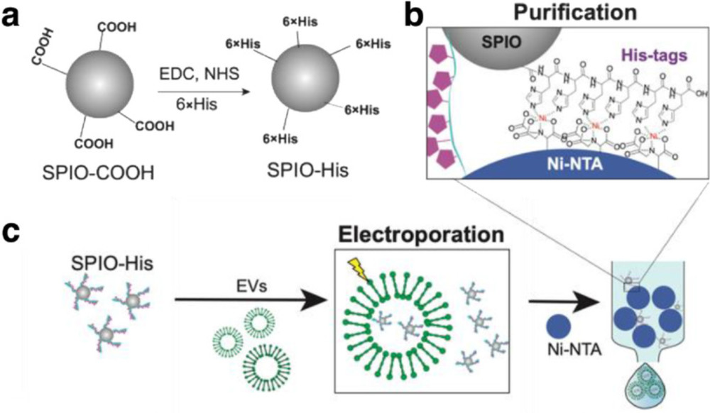 Figure 1. Schematic Overview of SPIO-His Functionalization and Exosome Loading. (Han Z, et al., 2021)
Our Advanced SPION Exosome Labeling Services
We deliver a fully characterized SPION labeling service designed around your specific imaging objective, including biodistribution mapping, tumor-homing validation, renal or hepatic clearance tracking, or theranostic agent development. Each project includes nanoparticle selection guidance, method optimization, and a complete analytical data package.
SPION Portfolio for Exosome Labeling
| SPION Type | Core Size (nm) | Contrast Mode | Primary Application | Key Feature |
|---|---|---|---|---|
| USPIO (dextran-coated) | 4-8 | T₂* / T₁ (low-field) | Passive cell-incubation labeling; lymph-node MRI | Long circulation; low immunogenicity |
| USPIO (PEG-coated) | 5-10 | T₂* / T₁ (low-field) | Systemic-injection EV tracking; renal clearance studies | Reduced opsonization; prolonged half-life |
| SPIO (carboxylated) | 15-30 | T₂ / T₂* | Electroporation loading; liver/spleen accumulation | High r₂; amenable to surface functionalization |
| SPIO (amine-functionalized) | 20-50 | T₂ / T₂* | Surface conjugation; targeting-ligand coupling | Covalent attachment to EV proteins |
| ES-SPION (citrate-stabilized) | 2-5 | T₁ / T₂ dual-mode | Theranostic exosome engineering; T₁-weighted angiography | Positive contrast capability; low magnetic artifacts |
| SPIO-Cy7 (dual-modal) | 15-30 | T₂* + NIR fluorescence | MPI-FLI bimodal tracking; ex vivo validation | Orthogonal detection: anatomical + optical |
| SPIO-His (histidine-tagged) | 15-20 | T₂* | Electroporation with Ni-NTA purification | Purifiable; >95% labeling efficiency |
We also source and validate custom SPION formulations upon request, including silica-coated, gold-core–shell, and manganese-doped iron oxide variants.
Labeling Strategies Available
We offer optimized, application-driven SPION exosome labeling workflows designed for MRI tracking, downstream purification, and preclinical translation:
- Electroporation loading (high-efficiency internal labeling): Parameter-optimized delivery of SPIONs into exosomes with controlled pulse conditions, followed by magnetic separation and removal of free nanoparticles.
- Parental cell incubation (physiological loading): SPION uptake by producer cells and natural packaging into exosomes, with EV isolation via ultracentrifugation/SEC and validation of intravesicular loading.
- Surface conjugation (targeted functional labeling): Chemical coupling of functionalized SPIONs to exosome membranes for enhanced signal stability and optional targeting modification.
- Dual-modality labeling (MRI + fluorescence): Integration of SPION MRI contrast with fluorescent reporters for complementary in vivo and in vitro tracking.
- Magnetic enrichment services: High-purity isolation of SPION-labeled exosomes and optional magnet-assisted targeting validation in experimental systems.
- Custom protocol development: Tailored workflows for specific exosome sources, SPION chemistries, and MRI platform, optimized for reproducible imaging performance.
Standard Workflow
- Study Design & Imaging Setup: Define MRI field strength, target tissue, and required signal sensitivity.
- Exosome Input QC: Verify EV size, concentration, and markers (NTA, CD63/CD81/CD9).
- Labeling Strategy Selection & Optimization: Choose SPION loading method (electroporation, co-incubation, or conjugation) and optimize key parameters.
- SPION Loading: Perform controlled exosome labeling under validated conditions.
- Purification & QC: Remove free SPIONs and assess iron content, integrity, and relaxivity (ICP-MS, NTA, TEM, r₁/r₂).
- Delivery & Imaging Report: Provide labeled exosomes with QC data and MRI guidance parameters.
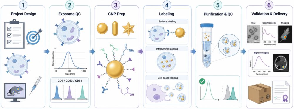
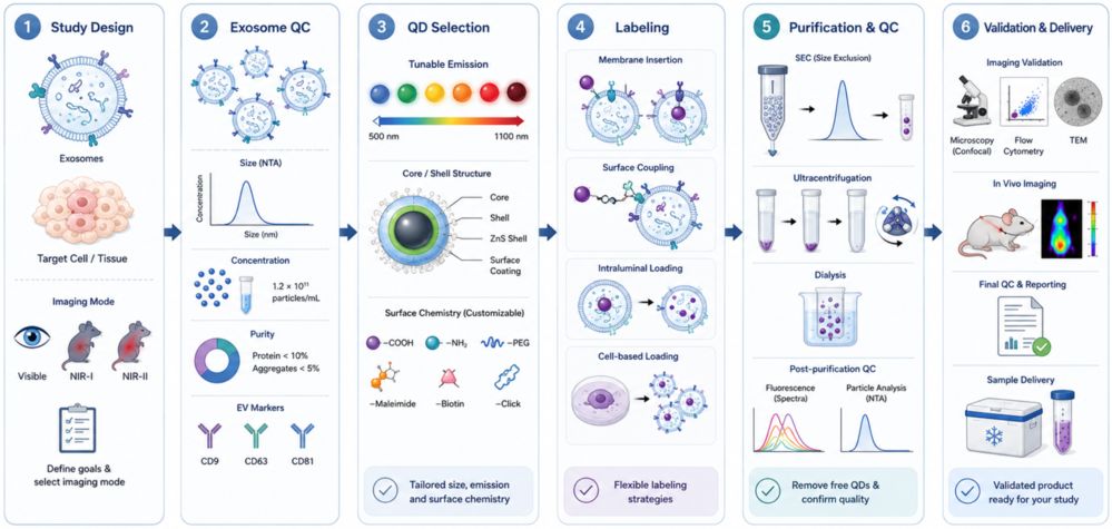
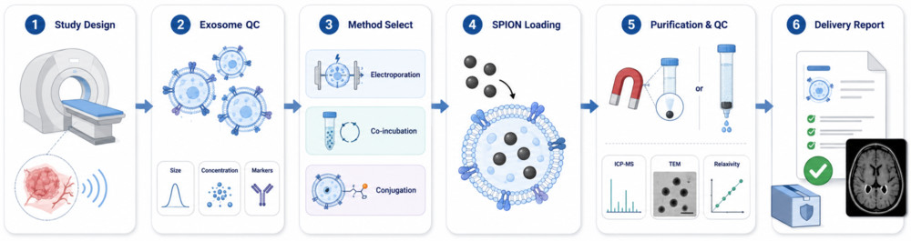 Figure 2. SPION Exosome MRI Labeling Workflow. (Creative Biostructure)
Quality Control and Characterization Standards
Each SPION-labeled exosome batch undergoes a streamlined, MRI-focused QC workflow to ensure imaging performance, purity, and reproducibility:
- Iron loading quantification: ICP-MS measurement of Fe content normalized to protein or particle number.
- MRI performance validation: r₁/r₂ relaxivity profiling at 1.5 T and/or 7 T for imaging compatibility.
- Size & concentration analysis: NTA-based particle counting with post-labeling aggregation assessment (DLS as needed).
- Structural confirmation: TEM imaging to verify vesicle integrity and SPION incorporation.
- Purity & identity markers: Tetraspanins (CD63/CD81/CD9) and contaminant markers (e.g., Calnexin) by Western blot or nano-flow cytometry.
- Free SPION removal check: Verification using matched controls and post-purification comparison assays.
All datasets are consolidated into a publication-ready QC report, with raw analytical files available upon request for regulatory or manuscript support.
Applications of SPION-Labeled Exosomes (Research Use Only)
- In vivo biodistribution: MRI-based whole-body and organ tracking with quantitative iron readouts.
- Tumor targeting: Longitudinal monitoring of exosome homing and retention in tumor models.
- Organ clearance: Liver, spleen, and kidney uptake and elimination profiling via ROI analysis.
- Lymphatic tracking: Non-invasive visualization of lymph node trafficking.
- Theranostics: Linking MRI-detected delivery sites with therapeutic payload response.
- Regenerative models: Tracking stem cell-derived exosomes in injury repair (heart, brain, kidney).
What Deliverables Will You Receive
| Deliverable | Description |
|---|---|
| SPION-Exosome Product | Sterile, MRI-ready EVs with defined iron loading and customized dose/concentration. |
| Iron Content Report | ICP-MS-based Fe quantification normalized to protein or particle count. |
| Relaxivity Data | r₁/r₂ values at specified field strength with key performance metrics. |
| QC Package | Core characterization: size (NTA/DLS), morphology (TEM), charge (zeta), EV markers. |
| Purity Controls | Free-SPION removal validation with matched controls for imaging baseline. |
| MRI Protocol Guide | Recommended sequences and parameters for T₂/T₂* exosome imaging. |
| Optional Add-ons | Stability test, uptake assay, MPI compatibility, and scale-up support. |
How to Get Started
We accommodate exosomes isolated by the client or provide end-to-end exosome isolation-plus-labeling packages:
- Submit your project brief: exosome source (cell line, biofluid, tissue), target imaging modality (MRI field strength, PET-MRI, MPI), and anticipated dose per animal.
- We evaluate feasibility, recommend SPION type and loading strategy, and deliver a technical proposal with timeline and pricing.
- Upon approval, pilot labeling is executed and QC data are reviewed with the client before full-scale production.
Why Choose Creative Biostructure for SPION Exosome Labeling
- Expert EV-nanoparticle engineering: Proven experience across electroporation, co-incubation, and covalent SPION loading strategies.
- Batch-specific MRI validation: r₁/r₂ relaxivity measured per batch at your selected field strength with no generic assumptions.
- Reliable signal specificity: Free-SPION removal rigorously validated to minimize false-positive MRI signals.
- Integrated QC workflow: Key analyses (NTA, TEM, surface markers, iron content, relaxivity) delivered in a single standardized report.
- Scalable & customizable production: From pilot studies to preclinical batches with tailored SPION chemistry and labeling density.
Case Study
Case: MRI Tracking of SPION-Labeled EVs
Background
Real-time in vivo tracking of therapeutic exosomes is essential for evaluating biodistribution and targeting, yet remains technically challenging. This study applied SPION-based magnetic labeling to enable MRI visualization of stem cell-derived EVs.
Methods
- Labeling: SPIO loading via electroporation
- Source: iPSC-derived EVs
- Purification: Removal of free SPIO by affinity-based QC
- Models: Kidney and myocardial injury
- Analysis: MRI, TEM, DLS, histology
Results
- Successful magnetic labeling with stable EV-SPIO complexes
- Clear MRI contrast enabling in vivo EV tracking
- Targeted accumulation in injured tissues post-injection
- Low background signal after free-SPIO removal
- Functional relevance consistent with regenerative effects
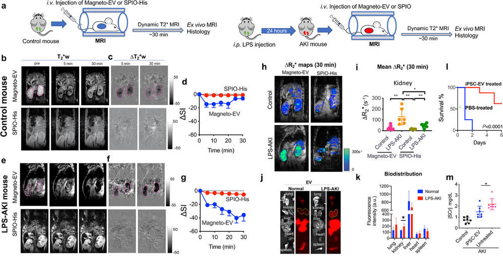 Figure 3. Representative T₂*-weighted MRI and quantitative maps showing in vivo distribution of SPION-labeled exosomes in normal and injured kidneys after systemic injection, with comparison of signal changes over time and organ-level biodistribution validation. (Han Z et al., 2021)
Conclusion
SPION labeling enables MRI-visible EV tracking with high specificity, supporting accurate biodistribution and preclinical functional studies.
Ready to track your exosomes non-invasively with SPION-enhanced MRI? Contact us to discuss nanoparticle selection, loading strategy, and a customized imaging-ready experimental plan.
References
- Busato A, Bonafede R, Bontempi P, et al. Magnetic resonance imaging of ultrasmall superparamagnetic iron oxide-labeled exosomes from stem cells: a new method to obtain labeled exosomes. International Journal of Nanomedicine. 2016: 2481-2490.
- Han Z, Liu S, Pei Y, et al. Highly efficient magnetic labelling allows MRI tracking of the homing of stem cell‐derived extracellular vesicles following systemic delivery. Journal of Extracellular Vesicles. 2021, 10(3): e12054.
Frequently Asked Questions
For any inquiries, our support team is ready to help you get technical support for your research and maximize your experience with Creative Biostructure.