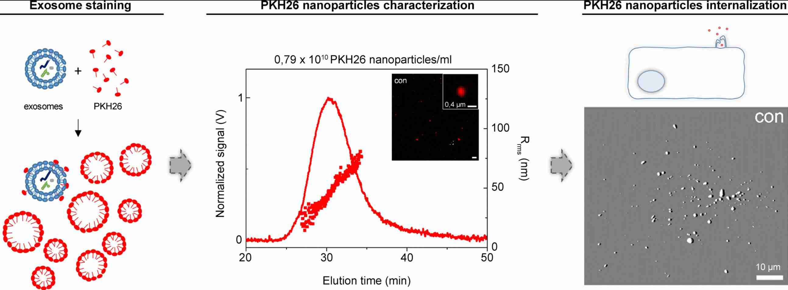
Exosome Labeling with Quantum Dots
Quantum dots (QDs) are 2-10 nm semiconductor nanocrystals with tunable emission, high photostability, and narrow spectra, enabling superior exosome labeling and long-term tracking versus organic dyes. Their resistance to photobleaching supports sustained in vivo imaging and multiplex detection with minimal spectral overlap for single-particle analysis. At Creative Biostructure, our QD exosome labeling service combines membrane insertion, covalent coupling, intraluminal loading, and metabolic incorporation with rigorous purification and characterization to deliver high-brightness, publication-ready exosomes for imaging, tracking, and biodistribution studies.
What Is Exosome Labeling with Quantum Dots
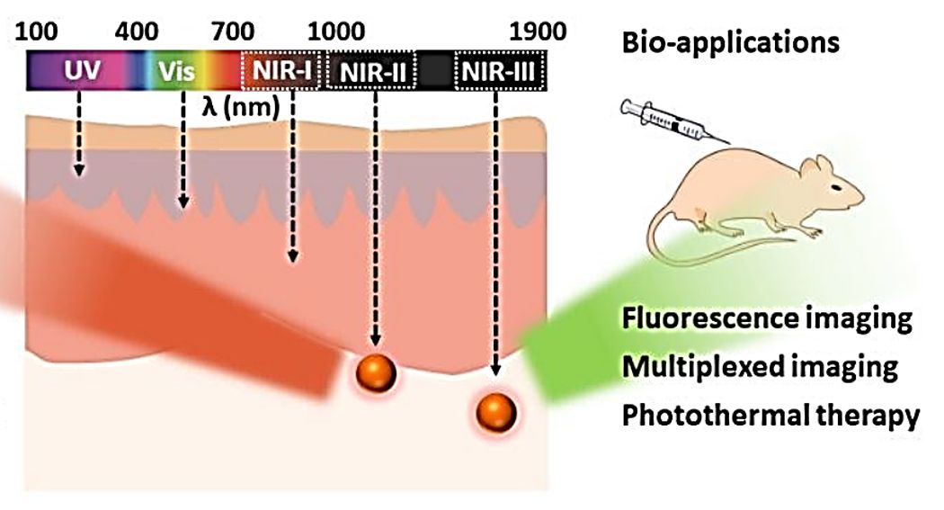
Exosome labeling with quantum dots (QDs) is the conjugation of semiconductor nanocrystals with extracellular vesicles (EVs) to generate highly bright, photostable fluorescent signals for sensitive exosome detection and tracking. Compared with organic dyes, QDs resist photobleaching and support long-term imaging of exosome uptake, trafficking, and subcellular localization. Their size-tunable emission spans from visible to the near-infrared II (NIR-II, 1000-1700 nm) window, enabling flexible selection for in vitro imaging or deep-tissue in vivo visualization.
QD-exosome labeling is typically achieved through four approaches:
(1) membrane insertion using hydrophobic QDs that integrate into the lipid bilayer;
(2) covalent coupling to surface proteins via EDC/NHS, thiol-maleimide, or click chemistry for stable surface tagging;
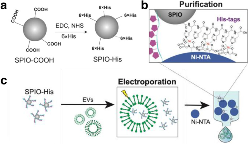
(3) intraluminal loading via electroporation, sonication, or permeabilization for protected internal tracking;
(4) producer-cell labeling, where QDs are taken up by donor cells and incorporated into secreted exosomes for biologically integrated, low-background labeling.
Our Advanced Quantum Dot Exosome Labeling Services
We offer customized quantum dot exosome labeling services tailored to imaging modality, study design, and workflow. Each project starts with technical consultation to select optimal QD properties, including emission, surface chemistry, and loading strategy, based on exosome source and detection requirements.
Quantum Dot Portfolio and Application Matrix
We stock validated quantum dots spanning the full UV-to-NIR-II spectral range with optimized surface chemistries for exosome conjugation:
| QD Type | Emission Range | Core/Shell Example | Primary Imaging Application | Key Advantages |
|---|---|---|---|---|
| Visible-range core/shell QDs | 450-650 nm | CdSe/ZnS | Multiplex confocal, super-resolution, flow cytometry | Narrow emission (FWHM 25-35 nm); exceptional brightness; well-established bioconjugation |
| NIR-I QDs | 650-900 nm | CdTe/CdSe, CuInS₂/ZnS | In vivo fluorescence, deep-tissue imaging | Reduced tissue autofluorescence; improved penetration depth |
| NIR-II QDs | 1000-1700 nm | Ag₂S, Ag₂Se, PbS/CdS | In vivo biodistribution, whole-body NIR-II imaging | Minimal tissue scattering; highest penetration; unprecedented SBR |
| Cadmium-free QDs | 500-750 nm | InP/ZnS, CuInS₂/ZnS | Biocompatible labeling, translational studies | Heavy-metal-free; reduced cytotoxicity; regulatory-friendly |
| Carbon / graphene QDs (CQDs/GQDs) | 400-600 nm | Carbon core, GQD sheets | Biosensing, chiral uptake studies, theranostics | Excellent biocompatibility; facile surface functionalization; low cost |
| Streptavidin/biofunctionalized QDs | Tunable (visible-NIR) | Pre-conjugated with SA, Protein A/G, or NHS ester | Immunolabeling, single-particle tracking | Ready-to-use; standardized protocols; rapid surface labeling |
| Perovskite QDs | 450-700 nm | CsPbX₃ (X = Cl, Br, I) | High-sensitivity biosensing, multiplex detection | Near-unity PLQY; ultra-narrow emission; emerging for EV applications |
Labeling Strategies We Offer
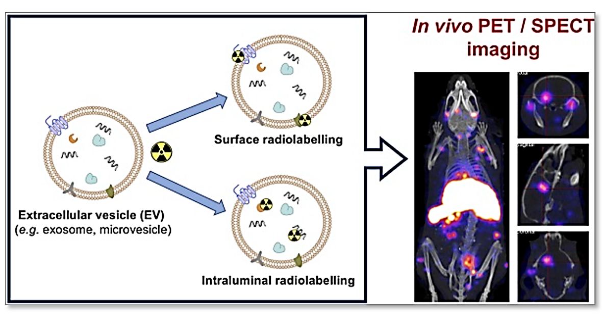
- Membrane insertion (hydrophobic QDs): Lipid-anchored QDs (e.g., alkyl, phospholipid, or cholesterol functionalization) passively integrate into the exosomal membrane under mild conditions. Suitable for rapid, non-invasive labeling in uptake, trafficking, and short-term tracking studies.
- Surface conjugation (covalent / immuno-based): Activated QDs (e.g., NHS, maleimide, or click chemistry handles) are covalently linked to exosomal surface proteins for stable, wash-resistant labeling. Optional targeting to EV markers (e.g., CD9/CD63/CD81) enables subpopulation-level detection and quantitative analysis.
- Intraluminal loading: QDs are encapsulated into vesicles via transient membrane permeabilization methods such as electroporation or sonication. This protects probes from the extracellular environment and supports long-term tracking and co-loading with experimental cargo.
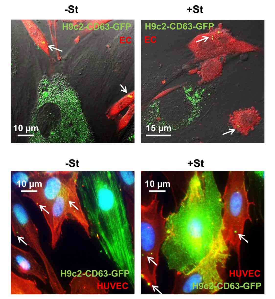
- Producer-cell loading: Functionalized QDs are internalized by donor cells and incorporated into secreted exosomes via the natural biogenesis pathway, enabling biologically integrated labeling with minimal surface modification and improved in vivo stability.
- Affinity-based labeling: Modular conjugation using biotin-streptavidin systems or QD-antibody constructs enables rapid and flexible labeling of engineered or native exosomes for immunodetection and fluorescence-based assays.
- Custom QD-EV design: QD properties (size, emission, surface chemistry, and ligand density) can be tailored to experimental needs, including multiplex imaging and hybrid probe integration, with full quality and stability validation.
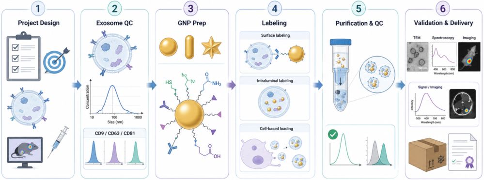
Standard Workflow
- Study design: Define exosome source, target, imaging mode (visible/NIR-I/NIR-II), and goal to select QD type, emission, and labeling strategy.
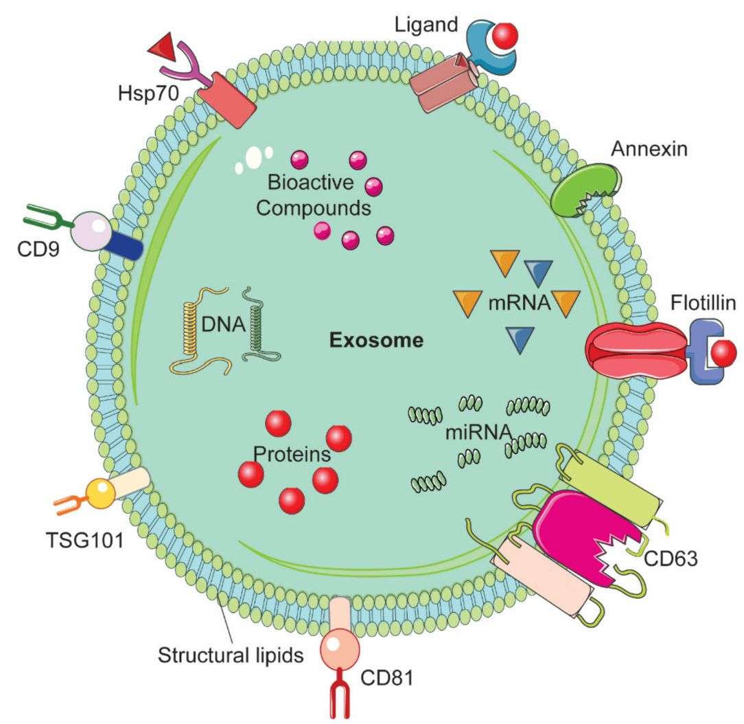
- Exosome QC: Assess size (NTA), concentration, purity, and EV markers (CD9/CD63/CD81).
- QD selection: Customize core/shell, emission, and surface chemistry per application needs.
- Labeling: Apply optimized QD-exosome conjugation (membrane insertion, covalent coupling, permeabilization, or cell loading).
- Purification & QC: Remove free QDs (SEC/ultracentrifugation/dialysis); verify labeling by fluorescence and particle analysis.
- Validation & delivery: Confirm via imaging/flow cytometry/TEM/in vivo assays; deliver with QC report and storage guidance.
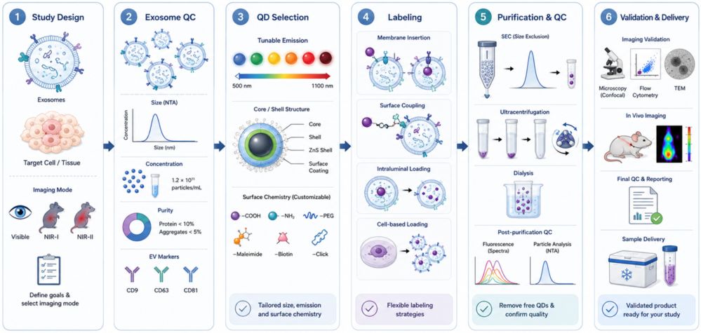 Figure 1. Quantum Dot Exosome Labeling Workflow. (Creative Biostructure)
Comprehensive Quality Control and Characterization
Each QD-labeled exosome batch undergoes comprehensive QC to verify particle integrity, labeling performance, purity, and functional suitability.
- Size & integrity: NTA and DLS analysis confirm particle size distribution and aggregation status before and after labeling.
- Labeling efficiency: Fluorescence analysis and nano-flow cytometry evaluate QD incorporation and signal intensity.
- Morphology & localization: Transmission electron microscopy (TEM) or Cryo-EM validates vesicle structure and QD association.
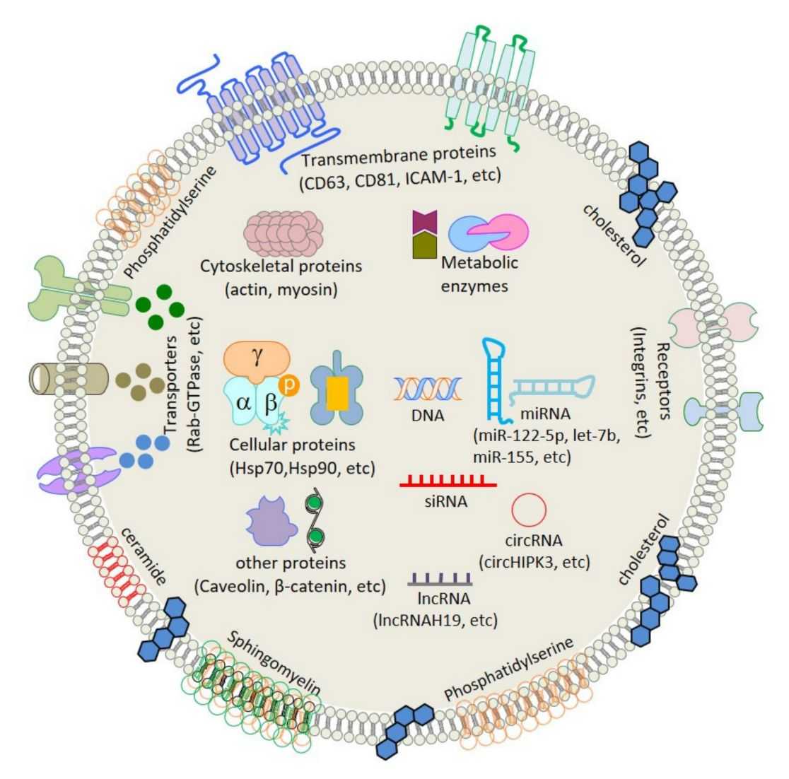
- EV identity: CD9/CD63/CD81 profiling confirms retention of key exosome markers after labeling.
- Optical performance: Emission spectra and photostability assays verify fluorescence characteristics.
- Purity assessment: Free QDs are evaluated and removed through optimized purification workflows with appropriate controls.
- Functional validation: Optional cellular uptake, multiplex imaging, and in vivo tracking assays are available based on project requirements.
All results are compiled into a publication-ready QC report with supporting data for downstream analysis and visualization.
Applications of Quantum Dot-Labeled Exosomes (Research Use Only)
QD-labeled exosomes provide bright, photostable, and multiplex-capable signals for advanced EV tracking and characterization in preclinical research.
- Multiplex exosome profiling: Enable simultaneous detection of distinct EV subpopulations using spectrally unique QDs for single-particle analysis and biomarker studies.
- Single-particle tracking: Support long-term visualization of individual exosomes to study cellular uptake, intracellular transport, and subcellular localization.
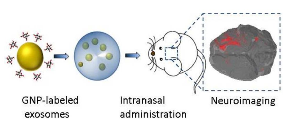
- In vivo biodistribution studies: NIR-I/NIR-II QD-labeled exosomes enable non-invasive tracking of tissue distribution, clearance, and targeting behavior in animal models.
- Cellular uptake & trafficking: Provide extended fluorescence imaging for monitoring exosome internalization and intracellular dynamics.
- Super-resolution imaging: High brightness and photostability make QDs suitable for nanoscale visualization of exosome interactions.
- Theranostic research: Facilitate simultaneous tracking of exosome-based cargo delivery and therapeutic responses in preclinical models.
- Biosensing & liquid biopsy research: Support sensitive exosome detection platforms for biomarker discovery and disease-related EV analysis.
What Deliverables Will You Receive
| Deliverable | Description |
|---|---|
| QD-labeled exosomes | Research-grade QD-conjugated exosomes in optimized storage buffer with defined concentration, aliquot size, and storage conditions (-80 °C) |
| Technical project report | QD selection rationale, conjugation strategy, purification workflow, yield summary, and recommended handling protocols |
| Multi-modal QC dataset | NTA, DLS, zeta potential, fluorescence emission/excitation spectra, photostability curves, TEM/cryo-EM micrographs, and nano-flow cytometry data |
| Matched controls | Unlabeled exosome control, free-QD control, and labeling-specificity validation data for quantitative comparison |
| Functional validation & scale guidance | Cellular uptake assay, multiplex imaging compatibility report, in vivo imaging pilot data, and scale-up recommendations |
How to Get Started
Project initiation is designed to be fast and streamlined. Clients may provide isolated exosomes, conditioned medium, or producer cells depending on the selected labeling strategy.
- Share requirements: Specify exosome source (cell line, biofluid, tissue), target application (imaging modality, tracking duration, multiplex requirements), and preferred QD composition if known.
- Feasibility review: Our team assesses compatibility, recommends the optimal QD type and conjugation strategy, and provides a detailed experimental plan with timeline and deliverables.
Why Choose Creative Biostructure
- Versatile QD options: Access a broad range of quantum dots with customizable emission, surface chemistry, and biocompatibility for diverse exosome imaging applications.
- Optimized labeling strategies: Select the most suitable QD-exosome labeling approach based on exosome source, imaging modality, and research objectives.
- Comprehensive QC: Each batch is evaluated for particle properties, labeling efficiency, EV identity, morphology, purity, and optical performance to ensure reliable results.
- End-to-end EV expertise: Our integrated capabilities in exosome engineering, QD functionalization, labeling, and imaging support projects from design to data delivery.
- Reproducible and scalable solutions: Standardized workflows with quality documentation enable consistent results for exploratory studies and preclinical research.
Case Study
Case: Quantum Dot Immunolabeling for Sensitive EV Detection
Background
Scatter-based nanoparticle tracking analysis cannot reliably distinguish EVs from similarly sized contaminants, while conventional fluorescent probes may lack sufficient brightness and photostability. This study developed a quantum dot-based immunolabeling method for selective detection and profiling of marker-positive EVs.
Methods
- EV sources: A549, THP-1, and EA.hy926 cell cultures
- Labeling strategy: Quantum dot-conjugated antibodies targeting CD9 and CD63
- Optimization: Probe concentration, incubation time, reaction volume, and removal of unbound QDs
- Analysis: Fluorescence and scatter nanoparticle tracking analysis, Western blot, SEM, and comparative fluorophore testing
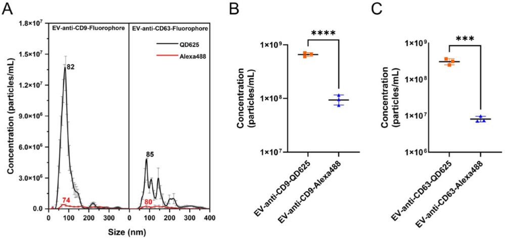 Figure 2. Fl-NTA comparison of A549-derived EVs labeled with QD625 or Alexa 488, showing higher particle detection with quantum dot labeling. (Ha E, et al., 2025)
Results
- Optimized labeling: QD concentration, incubation time, and reaction volume significantly affected labeling efficiency.
- Higher sensitivity: QD-labeled EVs produced substantially stronger fluorescence detection than EVs labeled with a conventional organic fluorophore.
- Small-EV detection: Fluorescence NTA detected marker-positive EV populations down to approximately 30 nm.
- Marker-specific profiling: CD9- and CD63-positive EV subpopulations showed distinct abundance and size distributions.
- Cross-cell-line analysis: The method revealed cell source-dependent differences in EV marker profiles across epithelial, endothelial, and monocytic cells.
Conclusion
Quantum dot immunolabeling improves the sensitivity and specificity of fluorescence-based EV analysis, supporting marker-selective detection, small-EV characterization, and comparative EV subpopulation profiling.
Ready to generate high-brightness quantum dot-labeled exosomes for your fluorescence imaging, single-particle tracking, multiplex profiling, or in vivo biodistribution project? Contact us to discuss QD composition, emission wavelength, conjugation strategy, and a customized experimental plan.
References
- Busato A, Bonafede R, Bontempi P, et al. Magnetic resonance imaging of ultrasmall superparamagnetic iron oxide-labeled exosomes from stem cells: a new method to obtain labeled exosomes. International Journal of Nanomedicine. 2016: 2481-2490.
- Ha E, Han Y, Kim M, et al. Quantum Dot‐Based Immunolabelling of Extracellular Vesicles and Detection Using Fluorescence‐Based Nanoparticle Tracking Analysis. Journal of Extracellular Biology. 2025, 4(7): e70072.
Frequently Asked Questions
For any inquiries, our support team is ready to help you get technical support for your research and maximize your experience with Creative Biostructure.