Structural Research of Polymerases
Polymerases play critical roles in living organisms. They are involved in the synthesis and repair of DNA and RNA. They catalyze nucleic acid polymerization reactions and link nucleic acid units (nucleotides) to form original DNA or RNA chains. Polymerases have highly conserved active sites, recognize and bind specific nucleotide sequences, and catalyze nucleic acid chain extension. Studying polymerases' structure is critical for understanding genome stability, disease mechanisms, and drug development. At the same time, by gaining insight into the structure and function of polymerases, the mechanisms of nucleic acid polymerization and the regulatory process can be revealed. This provides a basis for polymerase research and application in the life sciences.
Structural analysis of polymerase proteins can help determine how polymerases catalyze nucleic acid polymerization reactions, including the steps of substrate binding, catalytic transition state, and product release; reveal how they interact with molecules such as regulatory proteins, cofactors, and inhibitors to understand the regulatory mechanism; and provide important clues and a basis for designing inhibitors and drugs that target polymerases.
The first DNA polymerase was Pol I DNA from E. coli. The mechanism of the DNA polymerase reaction was validated using kinetic and X-ray structural data for the Klenow fragment (and its homologs from phage T7 and the thermophilic bacteria Thermus aquaticus and Bacillus stearothermophilus). DNA polymerases have conserved structures; the catalytic subunits vary very little in living cells of different species. This structural conservation and low variability may be due to the fact that these enzymes are very critical, even essential, for cellular processes.
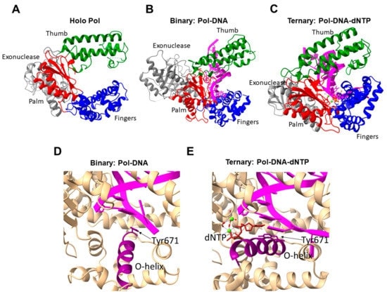 Figure 1. Three-dimensional structure of DNA polymerase I from E. coli: (Kuznetsova AA, et al., 2022)
| Protein | Organism | Method | Resolution | PDB Entry ID |
| TagF teichoic acid polymerase: Staphylococcus epidermidis | Staphylococcus epidermidis RP62A | X-ray diffraction | 2.70 Å | 3L7I |
| the Wall Teichoic Acid Polymerase TagF, H444N variant | Staphylococcus epidermidis RP62A | X-ray diffraction | 2.81 Å | 3L7J |
| the Wall Teichoic Acid Polymerase TagF, H444N + CDPG (15' soak) | Staphylococcus epidermidis RP62A | X-ray diffraction | 3.10 Å | 3L7K |
| he Wall Teichoic Acid Polymerase TagF, H444N + CDPG (30' soak) | Staphylococcus epidermidis RP62A | X-ray diffraction | 2.95 Å | 3L7L |
| the Wall Teichoic Acid Polymerase TagF, H548A | Staphylococcus epidermidis RP62A | X-ray diffraction | 2.85 Å | 3L7M |
Table 1. Structural research of polymerases.
Creative Biostructure offers services in polymerase structure research. We are a biotech company specializing in structural biology research. We provide services for crystallization screening and optimization of polymerases to obtain high quality crystals for X-ray crystallography studies. We also offer structure analysis and simulation services for polymerases to reveal their chemical and dynamic properties. This is done through X-ray crystallography, cryo-electron microscopy (Cryo-EM), and molecular dynamics simulations. With our advanced equipment and technology, we can help you better explore the mysteries of polymerases using structural biology methods and related services we offer. We can also provide the best structural biology services for life science research and drug development to help your project succeed. If you are interested in our services, please contact us for a more detailed description of our services.
Reference
- Kuznetsova AA, et al. Structural and molecular kinetic features of activities of dna polymerases. Int J Mol Sci. 2022. 23 (12).


