Structural Research of Photosystems
The primary energy supply for all higher life on earth is provided by the biological process of oxygenic photosynthesis, which converts light energy into chemical energy. This energy conversion is primarily a light-induced charge separation catalyzed based on two intramembrane protein complexes, photosystems I and II (PSI and PSII). Resolving the structures of PSI and PSII will improve the understanding of photosynthetic mechanisms and species evolution.
Advances in structural research of PSI
Cryo-electron microscopy is a favorable tool to research PS1. The structure at 3.8 Å resolution shows that PSI consists of 12 subunits, from PsaA to PsaX. PsaA and PsaB are the most essential parts, occupying most of the antenna system. Five C-terminal helices are located in the center of PSI and surround the electron transport chain, and six N-terminal helices are located on the side of the inner core. They are arranged as a trimer of dimers and coordinate the majority of the antenna pigments in PSI. Six smaller membrane-embedded proteins (PsaF, PsaI, PsaJ, PsaK, PsaL, and PsaX) surround the core to stabilize the antenna complex.
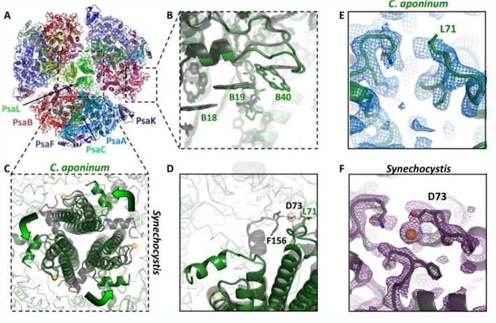 Figure 1. Trimeric structure of PSI from C. aponinum. (Dobson Z, et al., 2021)
Advances in structural research of PSII
PSII is a dimer in the photosynthetic membrane and consists of 17 subunits, from PsbA to PsbO. The researchers reconstructed an X-ray structural model of PSII at a resolution of 3.8 Å. They found that the central core of PSII contains an electron transfer chain consisting of a cluster of 2 × 5 transmembrane helices assigned to subunits D1 (PsbA) and D2 (PsbD). D1 and D2 are flanked by the antenna proteins D1 and D2 are flanked by the antenna proteins CP47 (PsbB) and CP43 (PsbC). These subunits all consist of six transmembrane helices, and comparison with PSI reveals a similar arrangement of transmembrane helices to the N-terminal portion of PsaA/PsaB.
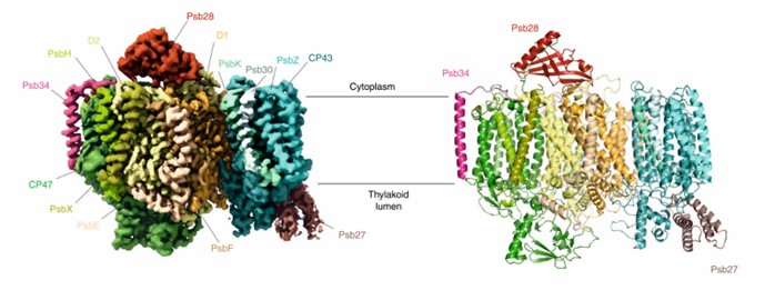 Figure 2. Crystal structure of PSII. (Zabret J, et al., 2021)
| Protein | Organism | Method | Resolution | PDB Entry ID |
| Photosystem I | Synechococcus elongatus | X-ray diffraction | 2.5 Å | 1JB0 |
| Photosystem I | Synechocystis sp. PCC 6803 substr. Kazusa | X-ray diffraction | 4 Å | 6HBQ |
| Trimeric Photosystem I | Cyanobacterium aponinum 0216 | Cryo-EM single particle analysis | 2.7 Å | 6VPV |
| PSI Monomer | Thermosynechococcus vestitus BP-1 | X-ray diffraction | 6.5 Å | 7BW2 |
| Photosystem I trimer | Synechocystis sp. PCC 6803 | X-ray diffraction | 2.501 Å | 5OY0 |
| Photosystem I in the presence of cytochrome c6 | Thermosynechococcus vestitus BP-1 | Cryo-EM single particle analysis | 2.85 Å | 6TRA |
| Photosystem I Acclimated to Far-red Light | Fischerella thermalis PCC 7521 | Cryo-EM single particle analysis | 3.19 Å | 6PNJ |
| Red-shifted photosystem I complex | Synechocystis sp. PCC 6803 substr. Kazusa | Cryo-EM single particle analysis | 3.1 Å | 6UZV |
| Far-red light utilizing photosystem I | Acaryochloris marina MBIC11017 | Cryo-EM single particle analysis | 2.5 Å | 7COY |
| Grafix PSI tetramer | Cyanophora paradoxa | Cryo-EM single particle analysis | 3.8 Å | 7DR2 |
| Tetrameric photosystem I | Nostoc sp. PCC 7120 = FACHB-418 | Cryo-EM single particle analysis | 2.37 Å | 6K61 |
| Wild-type PSI monomer1 | Cyanophora paradoxa | Cryo-EM single particle analysis | 3.3 Å | 7DR0 |
| Photosystem II | Synechococcus elongatus | X-ray diffraction | 3.8 Å | 1FE1 |
| Photosystem II | Thermosynechococcus vestitus | X-ray diffraction | 5.9 Å | 4IXR |
| Photosystem II | Thermosynechococcus vestitus | X-ray diffraction | 3 Å | 2AXT |
| I-substituted Photosystem II complex | Thermostichus vulcanus | X-ray diffraction | 4 Å | 3A0H |
| PSII-I prime (PSII with Psb28, and Psb34) | Thermosynechococcus vestitus BP-1 | Cryo-EM single particle analysis | 2.68 Å | 7NHQ |
| PSII-I (PSII with Psb27, Psb28, and Psb34) | Thermosynechococcus vestitus BP-1 | Cryo-EM single particle analysis | 2.72 Å | 7NHP |
| PSII intermediate Psb28-PSII complex | Thermostichus vulcanus | Cryo-EM single particle analysis | 3.14 Å | 7DXH |
| Photosystem II complex | Thermostichus vulcanus | X-ray diffraction | 1.9 Å | 3WU2 |
| Photosystem II depleted of the Mn4CaO5 cluster | Thermosynechococcus vestitus BP-1 | X-ray diffraction | 2.197 Å | 5MX2 |
| Photosystem II in the S1 state | Thermostichus vulcanus | X-ray diffraction | 2.35 Å | 7CJI |
| Photosystem II in the S2 state | Thermostichus vulcanus | X-ray diffraction | 2.4 Å | 7CJJ |
| Photosystem II in the dark S1 state | Thermostichus vulcanus | X-ray diffraction | 2.25 Å | 7COU |
| Monomeric photosystem II | Thermostichus vulcanus | Cryo-EM single particle analysis | 2.78 Å | 7EDA |
| PSII-M | Thermosynechococcus vestitus BP-1 | Cryo-EM single particle analysis | 2.66 Å | 7NHQ |
| Photosystem II, rows of dimers crystal packing | Thermosynechococcus vestitus | X-ray diffraction | 2.437 Å | 4PJ0 |
| PSII intermediate Psb28-RC47 | Thermostichus vulcanus | Cryo-EM single particle analysis | 3.14 Å | 7DXA |
| Photosystem II (1F state, dataset1) | Thermostichus vulcanus | X-ray diffraction | 2.15 Å | 6JLK |
| Photosystem II (2F state, dataset1) | Thermostichus vulcanus | X-ray diffraction | 2.15 Å | 6JLL |
| Photosystem II (1F state, dataset2) | Thermostichus vulcanus | X-ray diffraction | 2.4 Å | 6JLN |
| Photosystem II (2F state, dataset2) | Thermostichus vulcanus | X-ray diffraction | 2.4 Å | 6JLO |
| Photosystem II (3F state, dataset2) | Thermostichus vulcanus | X-ray diffraction | 2.5 Å | 6JLP |
Table 1. Structural research of photosystems.
Creative Biostructure provides high-quality structural analysis services for photosystems and other biomolecules. Our team of scientists has extensive experience in the use of cryo-electron microscopy (cryo-EM) and X-ray crystallography to provide accurate and reliable structural information. We can also tailor our services to meet the unique requirements of each research project, based on the specific requirements of clients. If you are interested in our services, please contact us. Our team will provide excellent service and support to ensure your satisfaction.
References
- Dobson Z, et al. The structure of photosystem I from a high-light-tolerant cyanobacteria. Elife. 2021.10: e67518.
- Zabret J, et al. Structural insights into photosystem II assembly. Nat Plants. 2021.7(4):524-538.
- Malavath T, et al. Structure and function of wild-type and subunit-depleted photosystem I in Synechocystis. Biochim Biophys Acta Bioenerg. 2018.1859(9):645-654.
- Fromme P, Grotjohann I. Structure of Photosystems I and II. Results Probl Cell Differ. 2008.45:33-72.


